In our fast-paced time, many diseases are getting younger. Increasingly, women in reproductive age are diagnosed with the dangerous size of uterine fibroids. Very patients experience shock when the gynecologist informs them of the diagnosis. In this article we will consider the danger of nodular myoma. Are there any ways to quickly cure ailment? Is it possible to avoid this disease and what is its treatment in case the inevitable happened.
From the article you will also learn how dangerous fibroids are during pregnancy and whether conception itself is possible in this case. Myomatous nodes are compared with the size of the fetus during pregnancy to determine the size of the tumor. A node is considered small up to 5-7 weeks, medium - up to 7-11, large - more than 12.
Is myoma dangerous?
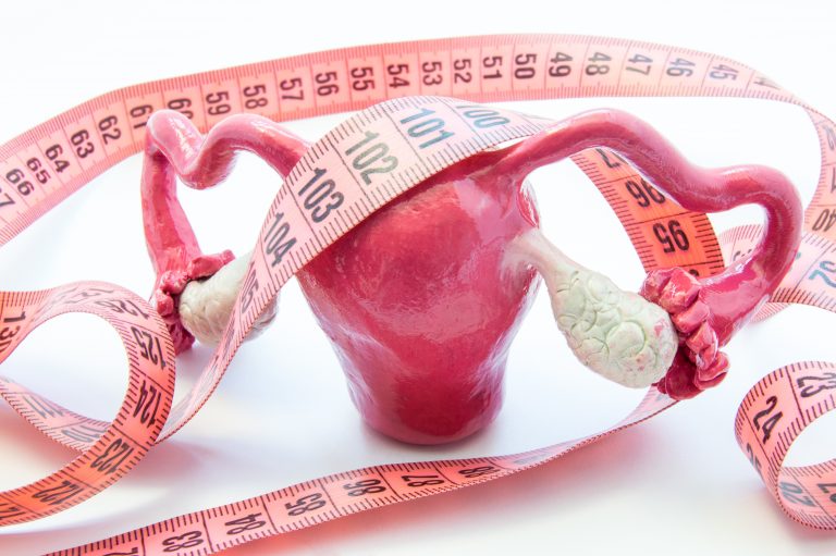
Is uterine fibroids dangerous for life? This disease occurs in 40% of young women. According to the modern concept, uterine fibroids are a genetically determined benign tumor that develops from smooth muscle cells of the uterus in the form of nodes. The neoplasm is a tangle of randomly interwoven smooth muscle fibers, which is visible to the doctor in the form of a round formation and is called the "myoma node" (myomatous node).
The main causes of the disease:
- bad habits;
- uterine injuries;
- hormonal disorders.
What is dangerous nodular myoma of the uterus? Diagnosis is required, during which it will be revealed whether the fibroid is a benign or malignant neoplasm. In the event that the tumor is the last, then uterine fibroids are dangerous for the patient's life. Surgery is required, and in some cases as soon as possible. Further therapy and prevention of relapse is carried out as prescribed by the attending physician.
Benign education
The nature of the neoplasm depends on the hormonal background and is often benign. Which does not alleviate the condition of the patient, as in some cases it threatens with serious complications. Uterine fibroids are life-threatening. According to statistics, myomatous nodes are found in 30% of women aged 25 - 45 years. They vary in shape, quantity, location. Some fibroids grow quickly, others slowly, but they all reach gigantic proportions, if you do not turn to a specialist in time!
The development of fibroids begins at the embryonic stage, when cells with altered genes are laid. In the subsequent of them, the growth of undifferentiated muscle cells begins, damaged and containing tumors in their structure. In the future, the development of fibroids can occur due to various factors.
Causes of neoplasm
The formation of the germ of myoma can occur at a later age as a result of the accumulation of gene mutations and the appearance of cells that give rise to tumor growth. About 10 years elapse from the moment the changes begin to develop the tumor.
The causes of the disease in 80% of patients are hereditary. Women in whom first-line relatives (mother, grandmothers, sisters) were ill with uterine fibroids are at 4 times more likely to develop this tumor than other women.
Uterine fibroids are the most common benign tumor of the female reproductive system. To date, no means have been invented that would prevent the development of tumors.
Who has a better chance of getting sick
Risk factors for the development of this disease or the progression of existing uterine fibroids:
- Hormonal changes.
- Irregular sex life or its absence.
- Early onset of menstruation.
- Lack of pregnancy and childbirth.
- Refusal (or impossibility) of breastfeeding.
- Violation of fat metabolism.
An alarming symptom that a woman should immediately pay attention to is more abundant menstrual flow, their duration. In the end, bleeding can be continuous for a month, continue outside the cycle (metrorrhagia), becoming more or less profuse.
Many women in this situation suffer and hope that everything, as it began by itself, will end, and do not seek help. In fact, you need to see a female doctor as soon as possible! Over time, if the patient does not seek help, this will lead to the development of anemia.

Women who are on the verge of menopause should be most attentive to their female health during this period, because at this age hormonal changes in the body occur and various diseases appear. This can be an impetus for the rapid growth of existing undiagnosed uterine fibroids nodes or their transition to the stage of malignant.
The effects of fibroids on women's health
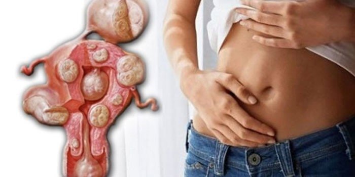
What is dangerous uterine fibroids? Myoma nodes grow very slowly, at first they are benign in nature, do not cause any pain and inconvenience, developing and taking up more and more space. If you do not start treating fibroids at the beginning of the disease, it becomes very large with heavy bleeding and this can result in infertility or degeneration of cells into a malignant tumor. Only 30% of patients older than 30 will experience unpleasant symptoms. Thus, at some point, the fibroids begin to really threaten the woman's life.
Uterine fibroids are very dangerous because at the very beginning the disease is asymptomatic. The node is detected accidentally during medical examination, on an ultrasound scan. It also happens that the disease has acquired a malignant form and nothing can be done already, that’s why myoma is dangerous.
Malignant neoplasm
Malignant transformation is influenced by:
- Smoking and alcohol abuse.
- Frequent tanning.
- Chronic diseases of the abdominal cavity.
- Warming up the pelvis.
What is the danger of nodular myoma? If it is not detected in time, then it grows, increases and occupies more and more space. With such dangerous dimensions, myoma causes concomitant diseases.
She begins to compress the abdominal organs that are located next to her (bladder, rectum, veins, etc.). This, in turn, leads to big problems with the organs of the urinary system. A woman has problems with urination, she has to go to the toilet more often, urinary incontinence is possible and cramping abdominal pains of a pressing nature appear. And because of pressure on the rectum, chronic constipation occurs.
It is necessary immediately as soon as the doctor diagnosed this disease, follow his recommendations. Thus, many problems can be avoided. Once a year, every woman needs to visit a gynecologist, do an ultrasound scan and identify serious diseases at the initial stage of development or make sure that they are absent. Any myoma is an unacceptable process that occurs in the uterus and requires immediate treatment, and if necessary, surgical intervention.
Uterine fibroids and pregnancy

All these changes occur in the female body in the reproductive period. Is myoma dangerous during pregnancy. A woman with a myomatous node may very well become pregnant. This is a difficult problem, since at the initial stage the disease is asymptomatic, and the woman does not feel or suspect anything about him.
A complication of pregnancy can occur at different times. In this period, most often there is an active growth of myomatous nodes, which is associated with hormonal changes in the body of a woman. They increase, soften, become more mobile.
Danger of development during pregnancy
Why is myoma dangerous? Large nodes deform the uterine cavity, creating a danger to the child, which leads to fetal hypoxia, the risk of miscarriage.
Serious complications include:
- necrosis of the uterine fibroids:
- placental insufficiency, when the placenta coincides with the location of the myomatous node;
- varicose veins as a result of the fact that large uterine fibroids compress veins;
- premature birth.
In the first trimester, the risk of miscarriage in women with uterine myoma is 47 - 50%.
Preservation of pregnancy with uterine myoma

In this case, it depends on the size of the node, on its location, age and individual characteristics of the woman’s body are also of great importance. It is impossible to identify the general tendency of delivery in the presence of fibroids, in each case, everything happens in different ways.
In the case of continued pregnancy, the expectant mother should regularly visit a gynecologist and carry out all the prescribed therapy. Usually, after childbirth, it is not a matter of preserving breastfeeding, the child has to be transferred to artificial nutrition with mixtures.
If complications arise, the doctor immediately hospitalizes the patient in the hospital, where treatment will continue. Violation of contractions during childbirth carries the danger of anomalies in labor. Contractions become not systematic, but attempts are weak and ineffective. This condition becomes life-threatening for the patient and the child, which leads to surgical delivery.
Modern diagnostic methods
Currently, the two main methods for the early diagnosis of fibroids are:
- Ultrasound.
- Radiation research methods.
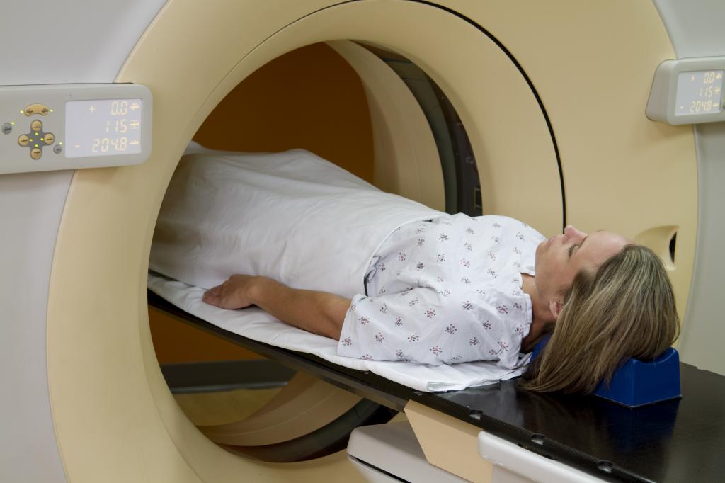
Over the years, the most accurate way to diagnose uterine disease and follow-up is the ultrasound method. It allows you to identify the danger of fibroids. On ultrasound, you can see small miamotous nodes (up to 1 cm in size), which cannot be detected by other methods in gynecological examination. The most optimal use of 3D-4D ultrasound, preferably color Doppler mapping. This will accurately determine the most dangerous sizes of uterine fibroids, its localization and interaction with surrounding tissues, as well as nearby blood flow.
It is also possible to combine ultrasound with echo hysterography. During the last procedure, a contrast fluid is injected, which will allow to differentiate fibroids from the uterine body, from polyps and endometrium, as well as to determine the location of the node, sizes and what danger it poses.
Radiation diagnostic methods include the use of computed or magnetic resonance imaging, multispiral computed tomography. These methods are not used so widely because they have a large radiation load on the body as a whole.
But they justified themselves in the diagnosis of large nodes, when the myoma is dangerous for the patient’s life, to clarify the nature of the location of the nodes, if it is not clearly visible on ultrasound, what is the cause of this pathology. They help determine the ratio of the tumor to related organs and systems, clearly define the boundaries of the formation, and also to find the vessels that feed this node, which can be used during embolization.
Classification of neoplasms by size
Which myoma is dangerous? Myoma can be classified by size:
- small myoma less than 2 cm;
- average fibroids from 2.1 cm to 6.9 cm;
- large fibroids from 6.9 cm to 10 cm;
- giant fibroids when the node reaches a size greater than 10 cm.
The size of the uterine fibroids becomes dangerous when the nodes reach average values.
The main methods of treatment
Identification of the disease in the early stages of development in the form of small nodes requires only observation. But what if the node begins to grow? The choice of method depends on the size of the body of the fibroids, the reproductive plans of the patient, concomitant diseases.
There are three treatment methods:
- Pharmacological.
- Surgical.
- Ray.
Most often, the patient, who has a medium-sized knot, does not plan to give birth and does not bother her, they are left under dynamic observation. This means that a woman should visit a gynecologist every 3-6 months for examination or ultrasound.
There is also a pharmacological method for treating uterine nodular fibroids. It aims to reduce the risk of bleeding and the amount of blood loss. The drug method can be a preoperative preparation in order to stabilize the growth of the node. Recently, one can often find the opinion that the treatment of uterine fibroids is possible using hormonal therapy. This method for a while allows you to either delay or completely move away from surgical treatment.
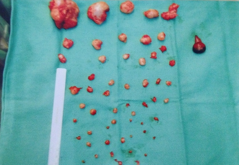
There are 3 methods of surgical treatment:
- removal of the uterus (hysterectomy);
- conservative myomectomy (allows you to remove the node, but at the same time save the uterus);
- hysteroresectoscopy (performed using high-tech equipment that allows you to control all manipulations inside the uterine cavity, which eliminates complications after surgery).
The choice of surgical method of treatment depends on the size of the node and its location, as well as on the skill of the surgeon. It is carried out only in specialized medical institutions equipped with appropriate equipment.
Radiation treatment methods include embolization of the uterine vessels and magnetic resonance FUS-ablation. These methods have a limitation to use, as they cannot be recommended to women planning a pregnancy due to the high risk of complications. The treatment method is selected only individually.