In the article, we will consider how the decoding of mammography of mammary glands is carried out.
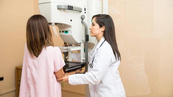
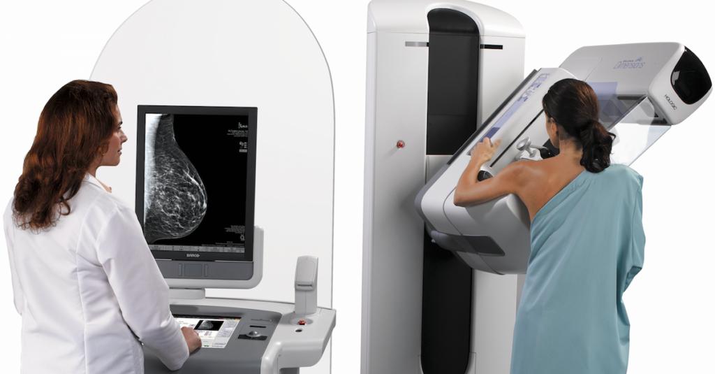
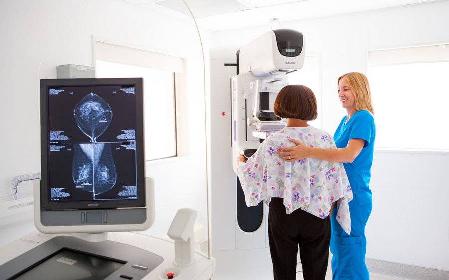
This is an X-ray examination. The doctor takes pictures in two projections, which is necessary for a better examination. When the results of a mammogram are ready, the specialist processes them, sometimes comparing them with previous studies. According to them, we can conclude about the presence or presence of any pathology.
So, what can the decoding of the results of mammography of the mammary glands reveal?
Possible pathologies
This diagnostic study is necessary to prematurely identify pathologies that are benign or malignant. Among them:
- Calcifications. They are an accumulation of calcium salts in the tissues of the mammary glands. Most often, this neoplasm indicates a developing oncological disease. Ordinary palpation does not allow to detect calcifications, so there is a need for mammography.
- Cysts They are a fairly common type of neoplasms filled with fluid. If a cyst is detected in the mammary gland, one should not panic, since such a pathology is not a sign of cancer.
- Fibroadenoma. It is a benign formation, prone to rapid development. Fibroadenomas are subject to surgical removal.
- Oncology - is a malignant neoplasm, characterized by uncontrolled growth. Cancer cells have the ability to invade adjacent cells and organs. An oncological neoplasm should be removed immediately.
After the patient is examined, the specialist receives x-rays of the mammary glands. Based on them, he determines whether there are any changes in the tissues.

Important factors to consider
In deciphering the results of mammary mammography and ultrasound, the doctor takes into account the following important factors:
- The presence of thickenings in the skin, calcifications.
- The presence of a variety of pathologies.
- Asymmetry (when only one of the mammary glands has seals).
It should be noted that it is impossible to diagnose cancer, relying solely on the results of a mammographic study. To determine the final diagnosis, the doctor recommends an additional examination. However, the results of the test can tell the specialist a lot.
Breast Mammography Decryption
Using an x-ray of the mammary glands, the doctor examines the condition of the lymph nodes, ducts, blood vessels, tissue texture. If there are no seals, the structure of the tissues in the chest is uniform, then we can judge the absence of pathological changes.
The ducts and capillaries in the images should be clearly visible. In the presence of violations of the structure of breast tissue, an increase in lymph nodes, the presence of pathology is diagnosed.
With the help of decoding mammography of mammary glands, it is not difficult for a specialist to determine the foci of development of the neoplasm, its quality, shape, size.
Categories
In accordance with accepted standards, the research results are divided into seven categories:

- Category 0. There is no necessary information in the image; additional studies are required. This category includes images during which the radiologist had doubts. Often, to assess the real state, images that were taken previously are used. As additional checks, an ultrasound examination, a mammogram in a different projection, and an enlarged view are prescribed.
- Category 1. In the tissues of the mammary glands, pathologies and compaction are absent. In this case, it is concluded that the woman is healthy. This indicator is considered to be the norm. This category includes images in which the mammary glands are symmetrical, in their structure there are no lumps, deformations, distortions of the structure, suspicious calcification.
- Category 2. There is an education that has a benign character, with no oncological symptoms. To describe the specialist uses obviously benign changes: fibroadenoma, enlarged lymph nodes, calcification. Obtaining such a result is guaranteed to indicate the absence of cancer.
- Category 3 in the decoding of the results of mammography of the mammary glands means that there is a neoplasm of a benign nature that requires additional research. The next examination should be done after six months. In addition, a woman needs to register with a mammologist. Approximately 98% of the detected formations are benign.
- Category 4. Suspicious seals were detected in the structure of the mammary gland. Most often, the risk of developing a cancer is extremely low. However, a woman is recommended to undergo a biopsy. There are 3 levels of cancer suspicion in this category: low (4A), intermediate (4B), moderate (4C).
- Category 5. Suspicious tumors are present in the structure of the mammary glands. In this case, a high probability of detecting a malignant neoplasm. To establish a final diagnosis, a biopsy is required.
- Category 6. A previously diagnosed oncology is detected in the structure of breast tissue. In this case, a mammographic study is used to evaluate the therapy performed and to control the growth of the malignant tumor.
If a specialist diagnoses the likelihood of developing cancer, panic is not worth it. It often happens that the indicators are normal, the decoding in the results of mammography of the mammary glands is incorrect.
False negative and false positive results
If the outcome of the examination shows the likelihood of the presence of cancer in the breast, the doctor recommends an additional check. It should be noted that mammography does not always give unambiguous and correct results.
If the specialist has at least the slightest suspicion of cancer, he sends the patient to additional studies. In the case when the diagnosis of the mammologist does not find confirmation, we are talking about a false-positive result of mammography. The woman in this case is recognized as healthy.

What is this fraught with
It is worth noting that such results negatively affect the physical and mental state of the patient. Most often, a woman, learning about the likelihood of the presence of a tumor, immediately begins to feel worse. In addition, a false-positive result implies further examination and, as a result, financial costs.
What mammography shows is important to determine in advance.
There are situations when the pictures reflect the normal state of the mammary glands, and after some time a woman is diagnosed with advanced cancer. In this case, the results of mammography are false negative.
According to statistics, cancer is not detected precisely for this reason in about 20% of patients. Typically, this situation occurs with young women. The structure of their mammary glands is more dense than that of older patients.

Factors Affecting Result
A false negative breast mammography result can be obtained due to several factors:
- The neoplasm has small dimensions.
- The doctor who performed the examination is inexperienced or incompetent.
- In the body of a woman, an increased level of sex hormones is noted.
- Malignant neoplasm grows dynamically.
Such a result is dangerous because a woman can put off a visit to a mammologist even if there are obvious symptoms of oncology. Such an approach to your health often leads to death. It is important to remember that only by decoding the mammography of the mammary glands cancer cannot be judged. If undesirable symptoms are present, a woman should immediately consult a doctor for advice.
Signs of Oncology
Symptoms arising from breast oncology depend on the stage of development and can be expressed by the occurrence of general weakness, sudden unreasonable weight fluctuations, changes in the shape of the breast, discharge from the nipple. A decrease in the size of the areola, deformation of the nipple, its retraction, and an increase in regional lymph nodes can also be observed.
Conclusion on the condition of the mammary glands
After a mammography, the mammologist will also have to evaluate the density of the mammary glands in the patient. In accordance with the generally accepted classification, 4 groups are distinguished:
- The predominance of adipose tissue. A certain amount of glandular and fibrous tissue is present in the structure of the mammary gland. The likelihood of developing a neoplasm is minimal.
- There are scattered portions of fibrous and glandular tissues.
- They have different densities. Detecting changes is difficult.
- Breast tissue is highly dense. Obtaining intelligible results using mammography is difficult. Oncological formations can mix with areas of normal tissues.
We examined how the decoding of mammography of mammary glands is carried out.