What does it mean if the doctor revealed hyperechoic inclusions in the uterus on ultrasound? Thanks to ultrasound scanning, a gynecological disease in a woman can be detected. This research method helps to study the necessary organs. To correctly understand the nature of the described formation, you need to familiarize yourself with the principles of operation of the ultrasonic device.
Diagnostic Features
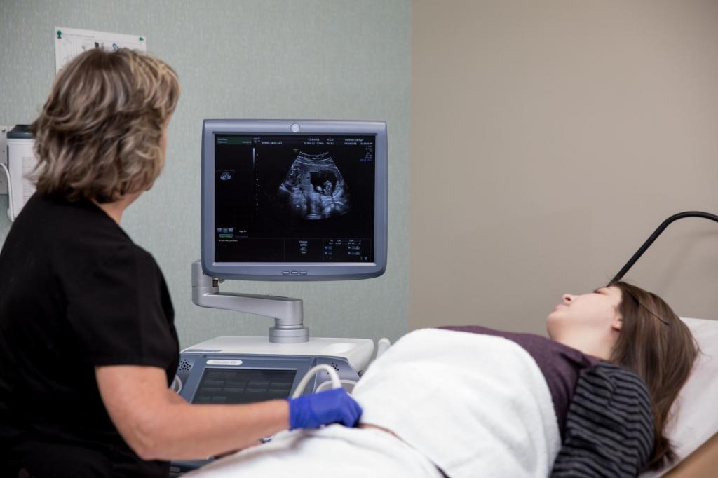
Echolocation is the basis of ultrasound. Due to the ability to reflect the sound waves of any structures, it is possible to identify any problems in the functioning of the pelvic organs. The piezocrystal, which is located in the sensor, produces an acoustic wave. In the process of conducting such diagnostics, various structures are scanned using the sound wave method of a special range. Such waves are able to propagate in biological education. The signal is sent to the tissue and reflected from it. Under these conditions, an image is created on the screen of the ultrasound machine. In the process of ultrasound, a specialist sees a black and white picture on the screen. In some cases, one ultrasound is not enough to diagnose a patient. Sometimes you should carry out a comprehensive diagnosis of the patient. With a high resistance of the biological material, relative to the signals, a clear picture is formed on the monitor. The denser the formation, the brighter the image. On the screen, the liquid is shown in black. There are several types of echogenicity. Among which are:
- normal
- increased;
- lowered.
The main varieties of inclusions

Given the degree of resistance of formations, the following types are distinguished:
- Hyperechogenic inclusions in the uterus. Characterized by high sound density. In the process of scanning, inclusions look like white or light spots.
- Hypoechoic variety. These structures have a low acoustic density compared to other tissues. In frequent cases, this indicates that an inflammatory process develops or edema has developed. On the monitor of the device, such formations appear as dark spots or black.
- Echogenic inclusions correspond to normal values, since the acoustic density is identical to the uterine tissue parameter. The object is shown in gray.
- Anechogenic is a fluid formation in which there is no echogenicity. It appears in a dark color. At another site, hyperechoic inclusions are presented in the form of bone tissue, cartilage compaction.
Using transabdominal and transvaginal ultrasound, you can fully evaluate the functioning of the uterus, identify the presence or absence of hyperechoic inclusions in the uterus, diseases of the pelvic organs. Thanks to regular medical examinations, the development of many pathological conditions can be prevented. As medical practice shows, often in the process of scanning patients find pathological formations. Under such conditions, it is important to determine the valency of the claimed seal. If a high acoustic density is detected, then hyperechoic inclusions in the uterus are examined.
Possible hyperechoic neoplasms
In the presence of hyperechoic formations in the organ, additional diagnostics should be carried out. Among the most common structures and inclusions that the doctor identifies during the study of the patient, there are:
- intrauterine device
- stagnation of blood in the uterus;
- inflamed suture after surgery;
- fibrous polyp;
- chronic endometritis;
- uterine fibroids;
- uterine wall rupture;
- calcium deposits;
- tumor;
- the presence of air in the uterus;
- the remainder of the fetus after abortion.
Hyper-echogenic inclusion in the uterus may indicate that the patient has formed a lipoma - a benign formation from adipose tissue. Pathology is often detected in older women. Not in all cases revealed hyperechoic inclusions in the uterus make it possible to diagnose the patient - it is important to carry out a complete medical diagnosis of the patient. If after an ultrasound the doctor has not received a sufficient amount of the necessary information, then magnetic resonance imaging is performed.
Features of inclusions in the uterus
Not in all cases, hyperechoic inclusions found in the uterine cavity indicate that serious diseases develop. In some cases, such inclusions are a normal state of the human body, but still many indicate a disease.
- Intrauterine contraceptives are one of the most popular methods of protection. After using an intrauterine contraceptive device, problems often arise in the work of the reproductive system of women, namely: hormonal failure, the development of the inflammatory process, perforation of the uterine wall. During scanning, the spiral is visualized as a direct hyperechoic structure.
- Chronic form of endometritis. Ultrasound shows a hyperechoic area with a clear border of up to 6 mm in size. Such an education has an irregular shape.
- Endometritis on an ultrasound scan. A fibrous polyp is clearly visible on the monitor, since the hyperechoic inclusion of the cervix consists of a dense fiber that perfectly reflects sound waves. The fibrous structure most often occurs in women aged, so an ultrasound scan is enough to make a diagnosis. Endometritis often resembles polyps of a fibrous nature - the disease differs only in that the image of the ultrasound machine reveals a rounded shape and clear contours.
- Myoma is a benign neoplasm that can trigger the development of bleeding. If timely therapy is not carried out, then the neoplasm will damage the uterine vessel. There is a nodular and diffuse form of the disease. With a diffuse form, high echogenicity is observed, since the uniformity and density of the affected tissue is violated.
- Hematometer. In the process of hematoma development, a neoplasm scan resembles an embryo membrane. It can be blood clots that remain after critical days.
- Tumor, lipoma, fibroids - detected in the process of ultrasound with the detection of echopositive structure.
An enlarged, heterogeneous uterus with hyperechoic inclusions is often diagnosed in women after childbirth. This may indicate that blood clots remained after childbirth. In any case, it is necessary to consult your doctor.
Reason for immediate medical attention
If the menstrual cycle is violated, bleeding occurs, pain in the pelvic organs often worries, then you need to visit a gynecologist and pass all the necessary tests for the doctor to make a diagnosis. In the process of development of many female diseases, vaginal discharge appears that has an unpleasant odor. In addition, there is irritation and pain in the pelvic organs. The pathological secret has an unusual color and a thick consistency.
Reasons for the appearance of an unusual secret in women

You should be aware that unusual discharge may also occur due to:
- the presence in the vagina of a foreign body;
- non-compliance with basic hygiene rules;
- allergies to drugs;
- wearing synthetic underwear.
Pain in the lower abdomen may be absent, so doctors recommend a regular thorough medical examination - this will help prevent uterine cancer. If bleeding has begun, you must immediately call an ambulance or visit a gynecologist.
Polyps in the uterus: symptoms

Small hyperechoic uterine inclusions can be detected in the process of ultrasound examination. Polyps are neoplasms of a benign nature. The main symptoms of the disease include:
- periodic pain in the lower abdomen;
- the presence of spotting in the middle of the cycle;
- brown discharge after sexual intercourse;
- sharp pain occurs if blood circulation in the polyps is impaired.
The causes of the disease
There are several reasons based on which the disease develops. These include:
- hormonal disbalance;
- excess weight;
- diabetes;
- liver disease.
Doctors advise to do an ultrasound of the uterus from time to time. Hyperachogenic inclusions are often diagnosed in women after 35 years of age - in frequent cases, they indicate that the disease develops. Due to age-related changes in the body, various neoplasms may appear. Thanks to gynecological examination, colposcopy, ultrasound examination, it is possible to detect the presence of pathology. Treatment can be either conservative or surgical. If you delay with going to the hospital, then serious health problems can arise. Hormonal drugs should be prescribed by a strictly attending physician.
Doctors Recommendations

The presence of point hyperechoic inclusions in the uterus may indicate that inflammation is developing. Under such conditions, it is necessary to repeat the diagnosis after a month and pass additional tests. Self-medication can only aggravate the course of pathology. In order to prevent the development of the disease, it is necessary:
- timely treat diseases that affect the regularity of the menstrual cycle;
- when a neuroendocrine syndrome occurs, consult a doctor;
- lead a healthy lifestyle - do not have abortions, do not smoke, eat right;
- to carry out effective therapy of the hyperplastic process in its initial detection.
Note to women

Hypo- and hyperechoic inclusions in the uterus can be diagnosed using ultrasound. Gynecologists recommend a thorough medical examination once every six months - this will help prevent the development of many pathologies. As medical practice shows, most diseases do not cause discomfort at the initial stage of development, therefore it is important to regularly visit a gynecologist. If inclusions are detected, then this is not always a sign of a serious pathology. But still it is better to get additional consultation with your doctor. Based on the diagnostic results obtained, the doctor will prescribe, if necessary, complex therapy. It is forbidden to self-medicate and apply folk recipes in order to improve well-being - this will only aggravate the course of the disease and harm the general state of health.