Focal length of the eye. What is it like?
Before starting the article, I turn to small photographers - stock up on fire extinguishers.
Go!
This time I will try to do without analogies of the eye with a camera and the brain with a computer. Why?
From the very first attempts to study the brain by humans, people have been looking for analogies to facilitate understanding / explanation of its work. For each era there were examples - a person compared the brain with the most complex device of his time:
- steam engines
- lamp technology,
- today these are computers,
- in future…
Let us turn to physiology textbooks to avoid unnecessary misconceptions.

In this figure, he added explanations for convenience.
Let's start with the ophthalmology guide.
The total refractive power of the entire optical conducting system of the eye is called physical refraction.
Diopters of all optical media of the eyeball:
- cornea ~ 43 diopters,
- front camera ~ 3 diopters,
- lens ~ 19-33 diopters,
- vitreous body ~ 6 diopters.
The front chamber is filled with aqueous humor - a liquid that is close to water in optical properties. (Remizov AN “Medical and biological physics” p. 384)
It must be understood that the first three media are collecting light, and the vitreous body scatters it, so when calculating we subtract this value.
The refractive power is calculated in diopters according to a simple formula from geometric optics:
D = Dr + Dp.k + Dhr-Dst.t. = 43 + 3 + 19-6 = 59 diopters
The value of the lens in this calculation is 19 diopters , since it corresponds to its refraction in a relaxed state when we look into the distance.
Next, we translate the diopters into millimeters:
F = 1 / D = 1/59 = 0.0169 m = 17 mm.
Conclusion: the focal length of the human eye is ~ 17 mm.
At the stage of studying the optical properties of the eye, we have a value of ~ 17 mm.
Quote - “Take the case where the average physical refraction (60,0 D) in the eyeball with the anteroposterior size of the average size (23 mm). It is easy to calculate that with a thickness of the cornea of about 1 mm, anterior chamber depth of about 3 mm and a distance from the anterior pole of the lens to the nodal point of 2 mm, from the latter to the retina, just 17 mm remains, which will ensure the focus of parallel rays in the central fossa of the yellow spot , as it coincides with the main focal length. ”
S.A. Rukhlova “Fundamentals of Ophthalmology” 2006
But, I think, someone will object - the focal length should be about 50mm!

Why should and why some think so ? To answer this question we will move further - into the visual cortex.
David Hubel and Thorsten Wiesel in their famous works on the physiology of vision established that the path retina-> LKT- > primary visual cortex has a topographic organization .
This tells us that the order in which the optic nerve fibers exit the retina is maintained in the cortex V1.
And R. Tutell was able to visualize this statement. To do this, he took a macaque, stuffed it with tranquilizers and for 45 minutes showed a target with three radial circles. The monkeys looked at the drawing with only one eye. Before this venture, the animal was injected with radioactive 2-deoxyglucose.
Since neurons feed exclusively on glucose, the most active cells can be easily tracked — they consume the most sugar.
After that, the macaques stretched their primary visual cortex, froze and showed radioactive marks.
The result is in the figure below.

The smallest circle in the center of the target on a topographic projection in the crust occupies an area quite a bit smaller than the area of the outer circle. In humans, this effect is even more pronounced - the central part of the visual field is projected onto larger areas in the cortex.
To facilitate understanding, the following figure was created:
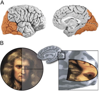
Here you can clearly see how the image is enlarged from the center of the retina.
I will emphasize that this is not an optical , but a cortical increase.
Summarize:
- focal length ~ 17 mm,
- coverage of the field of view of one eye horizontally 140 - 160˚,
- the image from the central part of the retina creates in the cortex a sensation (phenomenon) of an enlarged picture, although the optical projection is uniform.
Literature:
VVVit "The structure of the human visual system" 2003
E.A. Egorov “Ophthalmology” 2010
S.A. Rukhlova “Fundamentals of Ophthalmology” 2006
Novokhatsky A.G. Clinical Perimetry, 1973
David Hubel - Eye, Brain, Vision
Stephen Palmer - From Photons to Phenomenology
Baars B., Gage N. - “Brain, Cognition, Mind”
John Nicholls, A. Martin, B. Wallas, P. Fuchs - “From Neuron to Brain”
Michael Gazzaniga - “Who's in charge?”
References:
https://www.sciencedirect.com/science/article/pii/S089662730700774X
https://www.ncbi.nlm.nih.gov/books/NBK10944/
https://www.ncbi.nlm.nih.gov/pmc/articles/PMC5446894/
Go!
This time I will try to do without analogies of the eye with a camera and the brain with a computer. Why?
From the very first attempts to study the brain by humans, people have been looking for analogies to facilitate understanding / explanation of its work. For each era there were examples - a person compared the brain with the most complex device of his time:
- steam engines
- lamp technology,
- today these are computers,
- in future…
Let us turn to physiology textbooks to avoid unnecessary misconceptions.
Eye as an optical system
In this figure, he added explanations for convenience.
Let's start with the ophthalmology guide.
The total refractive power of the entire optical conducting system of the eye is called physical refraction.
Diopters of all optical media of the eyeball:
- cornea ~ 43 diopters,
- front camera ~ 3 diopters,
- lens ~ 19-33 diopters,
- vitreous body ~ 6 diopters.
The front chamber is filled with aqueous humor - a liquid that is close to water in optical properties. (Remizov AN “Medical and biological physics” p. 384)
It must be understood that the first three media are collecting light, and the vitreous body scatters it, so when calculating we subtract this value.
The refractive power is calculated in diopters according to a simple formula from geometric optics:
D = Dr + Dp.k + Dhr-Dst.t. = 43 + 3 + 19-6 = 59 diopters
The value of the lens in this calculation is 19 diopters , since it corresponds to its refraction in a relaxed state when we look into the distance.
Next, we translate the diopters into millimeters:
F = 1 / D = 1/59 = 0.0169 m = 17 mm.
Conclusion: the focal length of the human eye is ~ 17 mm.
At the stage of studying the optical properties of the eye, we have a value of ~ 17 mm.
Quote - “Take the case where the average physical refraction (60,0 D) in the eyeball with the anteroposterior size of the average size (23 mm). It is easy to calculate that with a thickness of the cornea of about 1 mm, anterior chamber depth of about 3 mm and a distance from the anterior pole of the lens to the nodal point of 2 mm, from the latter to the retina, just 17 mm remains, which will ensure the focus of parallel rays in the central fossa of the yellow spot , as it coincides with the main focal length. ”
S.A. Rukhlova “Fundamentals of Ophthalmology” 2006
But, I think, someone will object - the focal length should be about 50mm!
Why should and why some think so ? To answer this question we will move further - into the visual cortex.
Visual cortex
David Hubel and Thorsten Wiesel in their famous works on the physiology of vision established that the path retina-> LKT- > primary visual cortex has a topographic organization .
This tells us that the order in which the optic nerve fibers exit the retina is maintained in the cortex V1.
And R. Tutell was able to visualize this statement. To do this, he took a macaque, stuffed it with tranquilizers and for 45 minutes showed a target with three radial circles. The monkeys looked at the drawing with only one eye. Before this venture, the animal was injected with radioactive 2-deoxyglucose.
Since neurons feed exclusively on glucose, the most active cells can be easily tracked — they consume the most sugar.
After that, the macaques stretched their primary visual cortex, froze and showed radioactive marks.
The result is in the figure below.
The smallest circle in the center of the target on a topographic projection in the crust occupies an area quite a bit smaller than the area of the outer circle. In humans, this effect is even more pronounced - the central part of the visual field is projected onto larger areas in the cortex.
To facilitate understanding, the following figure was created:
Here you can clearly see how the image is enlarged from the center of the retina.
I will emphasize that this is not an optical , but a cortical increase.
Summarize:
- focal length ~ 17 mm,
- coverage of the field of view of one eye horizontally 140 - 160˚,
- the image from the central part of the retina creates in the cortex a sensation (phenomenon) of an enlarged picture, although the optical projection is uniform.
Literature:
VVVit "The structure of the human visual system" 2003
E.A. Egorov “Ophthalmology” 2010
S.A. Rukhlova “Fundamentals of Ophthalmology” 2006
Novokhatsky A.G. Clinical Perimetry, 1973
David Hubel - Eye, Brain, Vision
Stephen Palmer - From Photons to Phenomenology
Baars B., Gage N. - “Brain, Cognition, Mind”
John Nicholls, A. Martin, B. Wallas, P. Fuchs - “From Neuron to Brain”
Michael Gazzaniga - “Who's in charge?”
References:
https://www.sciencedirect.com/science/article/pii/S089662730700774X
https://www.ncbi.nlm.nih.gov/books/NBK10944/
https://www.ncbi.nlm.nih.gov/pmc/articles/PMC5446894/
All Articles