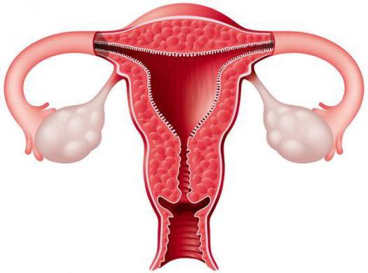
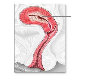
Endometrial adenomatosis is called atypical (focal or diffuse) endometrial hyperplasia, which, in fact, is a precancerous condition.
A precancerous process is a certain pathology, which with varying degrees of probability can go into cancer. The precancerous hyperplastic process has the possibility of reverse development, only 10% really turns into oncology. Uterine adenomatosis should be taken very seriously by doctors.
Disease Description

Hormonal dysfunction is directly related to hyperplastic processes in the endometrium. In this case, there are often uterine bleeding and infertility. They appear for the reason that there is hyperestrogenism. Excessive estrogen in the endometrium leads to quantitative and qualitative structural changes, which provokes the growth and thickening of its internal structures. So there is adenomatosis of the cervix.
Hyperplastic processes are of several types, depending on the type of cells that realize these processes in the body:
- glandular hyperplasia;
- diffuse hyperplasia;
- focal hyperplasia.
Let's consider each of them in more detail.

Glandular hyperplasia
When glandular structures increase, glandular endometrial hyperplasia develops. Sometimes this leads to cystic-expanded formations in the glands of the glands, then glandular-cystic hyperplasia is diagnosed. Atypical cells appear and grow in the endometrium, which is characteristic of adenomatosis.
It is important to understand that in case of impaired brain function, especially if the hypothalamus suffers, as well as weakened immunity and metabolic syndrome , cancer occurs in the case of glandular hyperplasia. Moreover, regardless of age.
Diffuse hyperplasia
In some cases, the spread of hyperplastic processes occurs along the entire surface of the endometrium, then specialists identify diffuse hyperplasia. That is, diffuse hyperplastic process leads to diffuse adenomatosis.
Focal hyperplasia

In addition, there is a focal form of hyperplasia. The growth of endometrioid tissue occurs in a limited area. Then this outgrowth disappears into the uterine cavity, which becomes similar to a polyp. Focal adenomatosis is a polyp in which there are atypical cells.
Uterine adenomatosis is treated mainly in a surgical way. The further forecast is determined by several factors:
- age of the patient;
- the nature of hormonal disorders;
- concomitant neuroendocrine diseases;
- a state of immunity.
Some women are interested in the question of what is the difference between uterine adenomatosis and endometrial adenomatosis? After all, this is one and the same atypical process. The term "uterine adenomatosis" is not entirely correct, since atypia affects only the inner layer, which is the endometrium. And in the uterus itself there are several layers.
Fibrosis and adenomatosis

Fibrous adenomatosis as a diagnosis does not exist. Fibrosis is a pathology in which connective tissue grows, adenomatosis - glandular growth. A pathology of a mixed nature may also have what will be called fibrocystic hyperplasia.
Adenomatosis can be not only in the uterus. It happens in the mammary glands, but in fact these pathological processes are completely different. Breast adenomatosis is Reclus disease, when a benign formation of small-sized cysts occurs. We examined cervical adenomatosis. What it is has become clearer.
What are the causes of endometrial adenomatosis?
The causes of atypical cell transformation are the same factors that provoke hyperplastic processes in the endometrium. Reliable causes of adenomatosis are not known. Of course, the provoking factors are constantly being studied, but so far it is impossible to say for sure that this is precisely the trigger of the atypical process in the endometrium. But the more there are various adverse conditions, the more likely the development of pathology.
The first place among all the provoking factors of endometrial adenomatosis is hormonal failure. The neurohumoral regulation of the entire human body is disturbed. Estrogens and gestagens are involved in physiological cyclic changes in the uterus. First of all, thanks to estrogens, an increase in the internal mucous layer occurs. But the work of gestagens consists in timely stopping the growth of the endometrium and its rejection.
With an excessive amount of estrogen, the growth of the endometrium occurs uncontrollably. Hyperestrogenism can happen for various reasons:
- hormonal function of the ovaries is disturbed;
- anovulation occurs;
- the cycle becomes single-phase;
- there is endometrial hyperplasia.

With polycystic ovary anovulation is chronic. This is also a kind of provoking factor for the development of hyperplasia. If a woman takes hormonal drugs uncontrollably, then the hormonal background can suffer from this. This will start the hyperplastic process in the endometrium.
If there is simultaneously hyperestrogenism, extragenital pathology and neuroendocrine disorders in the body, the likelihood of developing adenomatosis increases. A woman with obesity and hypertension is susceptible to endometrial cancer 10 times more than one who has normal weight and blood pressure.
For what reasons can hyperestrogenism still develop? Often diseases of the liver and biliary tract lead to this pathology, since it is the liver that utilizes estrogens.
So, there is an uncontrolled growth of the inner layer of the uterus, which leads to the formation of atypical cells. This is the endometrial adenomatosis. What is the treatment for a cervical adenomatosis? About it further.
Signs of endometrial adenomatosis
As a rule, there are no obvious symptoms of adenomatosis, since atypical cells can only be detected in a laboratory way. First, a hyperplastic process is detected, after which it is necessary to clarify its nature.

There are some symptoms of hyperplasia that you should definitely pay attention to:
- the nature of the bleeding is changed - menstruation becomes abundant, blood appears outside the cycle;
- pain in the lower abdomen and lower back before and during menstruation;
- manifestation of the metabolic syndrome - excess weight, excessive male type hair, increased levels of insulin in the blood;
- fertility is impaired - it is impossible to conceive and bear a child;
- the presence of mastopathy;
- inflammation of the genitourinary system;
- pain during sexual intercourse, blood discharge after it.
Is uterine adenomatosis detected by ultrasound?
Ultrasound scanning determines the thickness and structure of the endometrium. The transvaginal probe does a good job with this study. What kind of hyperplastic process is observed - focal or diffuse - this scan will show. As a result, if diffuse hyperplasia is detected, then we can assume the presence of diffuse adenomatosis. It is impossible to visualize it with a sensor, since there are no distinguishing features.
Focal uterine adenomatosis is easy to detect, since it is visualized as a polyp. Although the nature of cellular changes can also not be detected. Atypia is not traceable by ultrasound scanning.
A scraping of the uterine mucosa is made, after which this material is sent for histological examination. This diagnostic method is very important for adenomatosis. We study the composition of the cell, its structural change, and also to what extent and severity it is atypical. If atypia is not detected, then this indicates a benign course of hyperplasia.
Often surgical curettage of the uterine cavity is performed, and then the resulting material is examined. This can help hysteroscopy for visual control during total evacuation of the uterine mucosa.

Uterine adenomatosis: treatment
The presence of adenomatosis in a woman can be the cause of infertility, but even with successful conception against the background of the disease, premature termination of pregnancy can occur.
Treatment primarily consists of the fact that the altered endometrium is removed mechanically. Thus, the source of pathological changes is surgically eliminated, in addition, scraping for histological examination is obtained. When the results are obtained, the treatment plan is determined depending on this.
Hormone therapy and surgery are individually prescribed. If the girl is young, then specialists are limited to treatment with hormonal drugs. A patient who is at an age close to menopause, together with hormone therapy, undergoes a radical surgical operation - removal of the uterus and appendages. This greatly reduces the likelihood of adenomatosis becoming cancer. You can save a woman’s life.
It is important to understand that early diagnosis of adenomatosis is most desirable, in which case the risk of oncology is minimal. Therefore, it is necessary to regularly visit a gynecologist, undergo a comprehensive examination, take all the necessary tests. We examined in this article adenomatosis of the uterine endometrium. Take care of your health!