Squeak of a cancer tumor: scientists of NUST “MISiS” have developed a laser ultrasound for the diagnosis of cancer
A group of scientists from NUST “MISiS” developed a universal system of optical-acoustic ultrasound based on the use of ultrasonic waves and laser radiation. It can be used to obtain images of internal pathologies, including the detection of small-sized tumors that are not fixed by conventional ultrasound. The results are published in the international scientific journal Photoacoustics .

It is interesting that the development was done by specialists from the laboratory with the name (attention!) “Laser-ultrasonic diagnostics of the structure and properties of rocks and heterogeneous structural materials”, which is headed by Prof. Alexander A. Karabutov, Ph.D.
It would seem - where are the miners and where is oncology?
However, the method of laser ultrasonic spectroscopy studied by laboratory specialists turned out to be so universal that the technologies created on this basis can be applied in a wide variety of fields.
We already wrote on “Habré” about a device created in the laboratory for detecting defects in aircraft parts 50 microns in size and launched into small-scale production. And now - a new development in the field of medical technology.
As the creators said, initially the system was made to visualize blood vessels. However, in the course of research it turned out that the apparatus is also applicable for examining hard tissues (for example, teeth) and for diagnosing oncological formations in soft tissues.
The fact is that standard ultrasound methods due to low contrast and image quality do not allow, for example, to distinguish a cancerous tumor from healthy tissues at an early stage. Confident recognition is only possible if the tumor size exceeds 1 cm.
Our development provides the doctor with not only standard ultrasound images, but also additional information about those tissues that are poorly distinguishable acoustically (using ultrasound), but at the same time have different absorption capacity. Cancerous tumors also belong to such tissues.
The whole system is based on a well-known physical phenomenon - the optical-acoustic effect. It consists in the following: laser radiation of a very short duration is absorbed in the irradiated object (in this case, living tissues of the body), which leads to rapid heating of the site of this object. Heating leads to the expansion of tissue matter and the corresponding excitation of ultrasonic waves. Thus, irradiation with short laser pulses leads to "vibration" of the tissue site and their emission of ultrasound. Roughly speaking, a living organ “squeaks” in ultra-high tones.
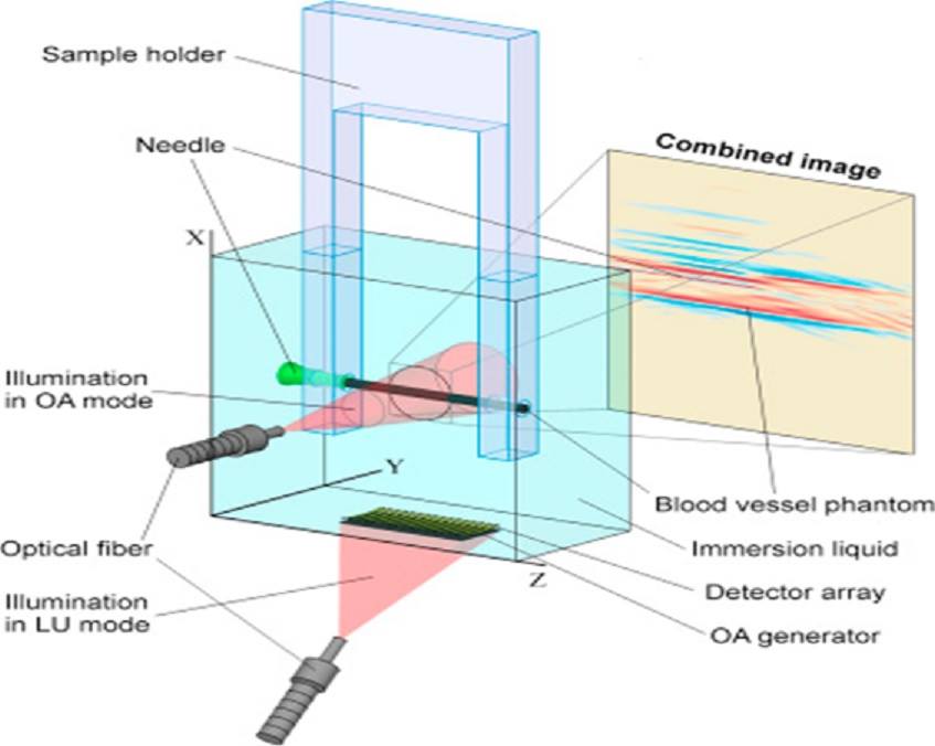
The scheme of the device
“In the installation, laser radiation is used to excite ultrasound in two modes. In the first, optical-acoustic, light is absorbed directly in the studied object (in this case, a small section of a blood vessel or tumor begins to “vibrate”). The waves excited in this way are recorded by a variety of receivers (a special acoustic antenna), and the signals from these elements are used in the future to build accurate images of the object that provide contrast in the absorption of light - explained one of the co-authors, engineer of the laboratory of laser ultrasonic methods of introscopic studies of NUST “MISiS” Vasily Zarubin .
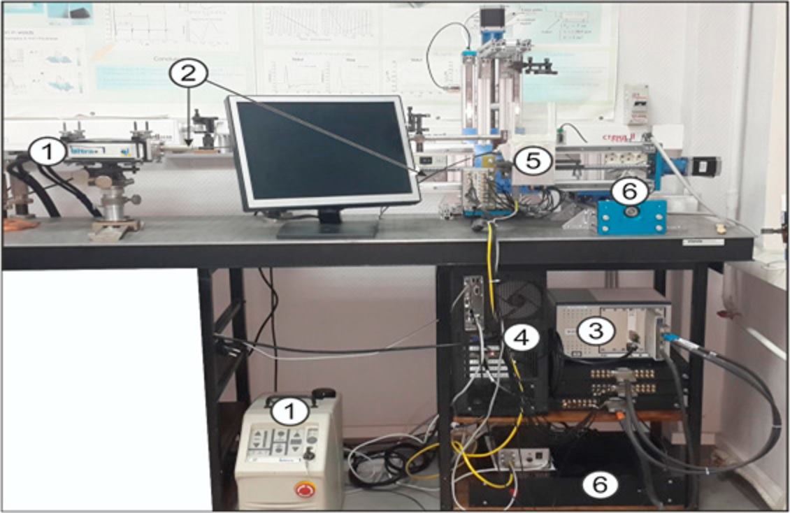
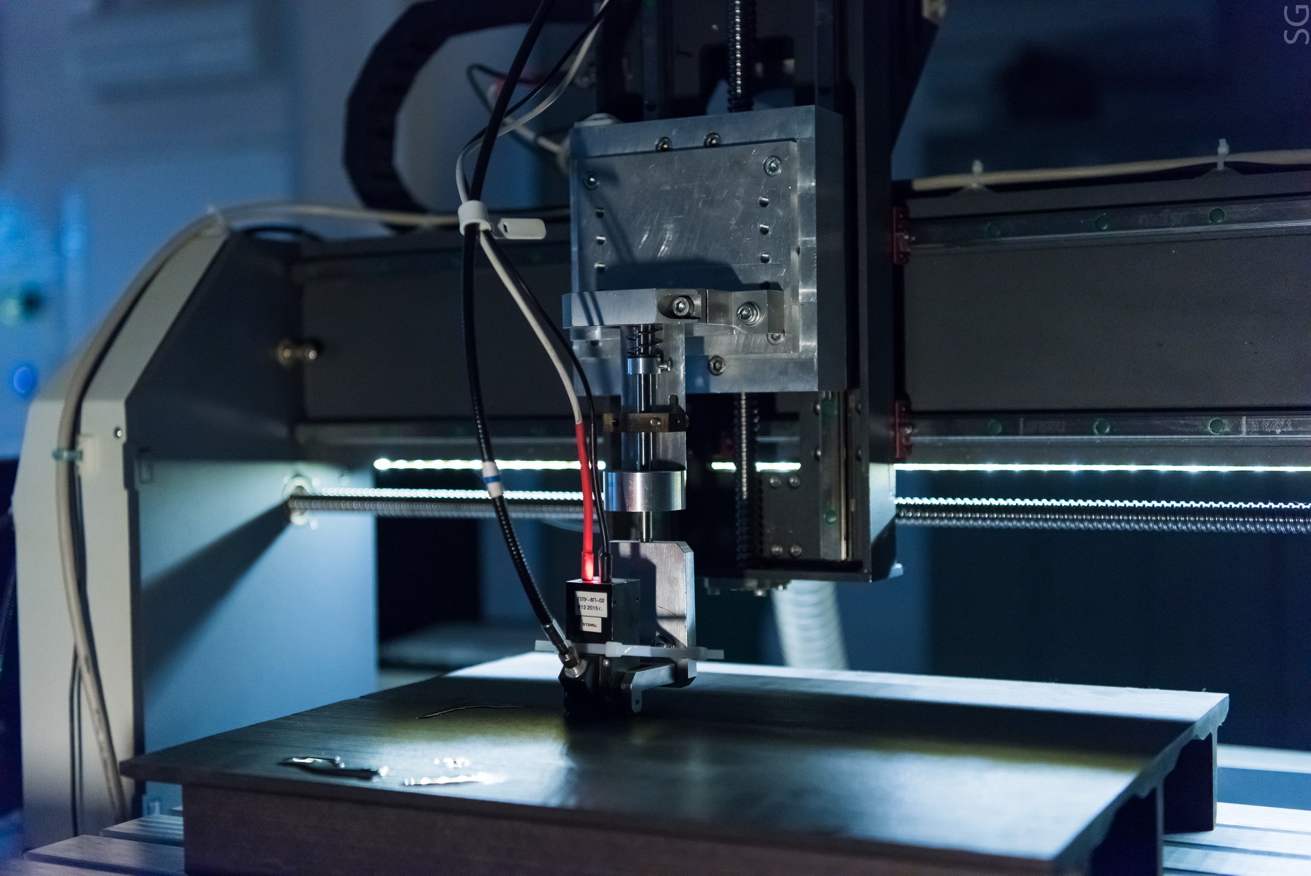
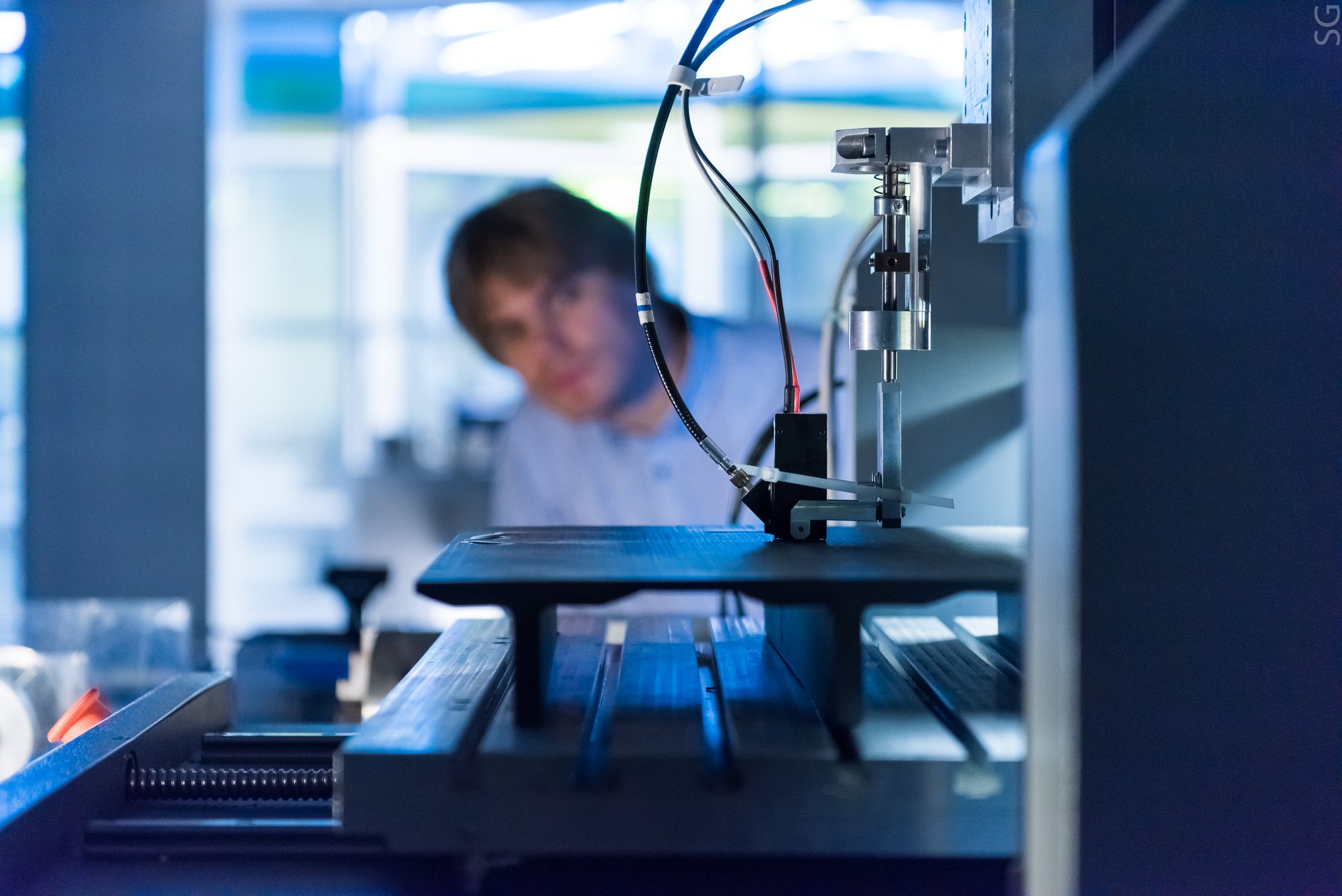
Photograph of the experimental setup: 1 - laser with a power source; 2 - fiber optic laser delivery system in LU mode; 3 - a system for collecting and processing experimental data; 4-RS; 5 - matrix of detectors; 6–3D positioning system.
- In the second, laser ultrasonic mode, the light is absorbed already in a special plate, and it begins to “vibrate”. The waves excited in it are used to study the object in a manner similar to standard ultrasound. In this case, ultrasonic waves are scattered by the inhomogeneities of the object, and are received by the same acoustic antenna. The signals from it are used to build the final laser ultrasound images. "
As a result, a device based on laser ultrasound makes it possible to detect a tumor at an early stage, with a size of less than a millimeter.
Currently, the research team is improving the characteristics of the experimental prototype of the system, as well as adapting it to specific tasks.
It is interesting that the development was done by specialists from the laboratory with the name (attention!) “Laser-ultrasonic diagnostics of the structure and properties of rocks and heterogeneous structural materials”, which is headed by Prof. Alexander A. Karabutov, Ph.D.
It would seem - where are the miners and where is oncology?
However, the method of laser ultrasonic spectroscopy studied by laboratory specialists turned out to be so universal that the technologies created on this basis can be applied in a wide variety of fields.
We already wrote on “Habré” about a device created in the laboratory for detecting defects in aircraft parts 50 microns in size and launched into small-scale production. And now - a new development in the field of medical technology.
As the creators said, initially the system was made to visualize blood vessels. However, in the course of research it turned out that the apparatus is also applicable for examining hard tissues (for example, teeth) and for diagnosing oncological formations in soft tissues.
The fact is that standard ultrasound methods due to low contrast and image quality do not allow, for example, to distinguish a cancerous tumor from healthy tissues at an early stage. Confident recognition is only possible if the tumor size exceeds 1 cm.
Our development provides the doctor with not only standard ultrasound images, but also additional information about those tissues that are poorly distinguishable acoustically (using ultrasound), but at the same time have different absorption capacity. Cancerous tumors also belong to such tissues.
The whole system is based on a well-known physical phenomenon - the optical-acoustic effect. It consists in the following: laser radiation of a very short duration is absorbed in the irradiated object (in this case, living tissues of the body), which leads to rapid heating of the site of this object. Heating leads to the expansion of tissue matter and the corresponding excitation of ultrasonic waves. Thus, irradiation with short laser pulses leads to "vibration" of the tissue site and their emission of ultrasound. Roughly speaking, a living organ “squeaks” in ultra-high tones.
The scheme of the device
“In the installation, laser radiation is used to excite ultrasound in two modes. In the first, optical-acoustic, light is absorbed directly in the studied object (in this case, a small section of a blood vessel or tumor begins to “vibrate”). The waves excited in this way are recorded by a variety of receivers (a special acoustic antenna), and the signals from these elements are used in the future to build accurate images of the object that provide contrast in the absorption of light - explained one of the co-authors, engineer of the laboratory of laser ultrasonic methods of introscopic studies of NUST “MISiS” Vasily Zarubin .
Photograph of the experimental setup: 1 - laser with a power source; 2 - fiber optic laser delivery system in LU mode; 3 - a system for collecting and processing experimental data; 4-RS; 5 - matrix of detectors; 6–3D positioning system.
- In the second, laser ultrasonic mode, the light is absorbed already in a special plate, and it begins to “vibrate”. The waves excited in it are used to study the object in a manner similar to standard ultrasound. In this case, ultrasonic waves are scattered by the inhomogeneities of the object, and are received by the same acoustic antenna. The signals from it are used to build the final laser ultrasound images. "
As a result, a device based on laser ultrasound makes it possible to detect a tumor at an early stage, with a size of less than a millimeter.
Currently, the research team is improving the characteristics of the experimental prototype of the system, as well as adapting it to specific tasks.
All Articles