Breast ultrasound with elastography - procedures that help to increase the range of application of ultrasound examination. Visual inspection of internal organs helps to get more detailed information. What is breast elastography? Breast sonoelastography - examination of the mammary glands for their density. If there are any malignant tumors in the chest, the device determines the densified areas and gives them a detailed description.
Description of procedure
Breast elastography - what is it? Tissues in the human body have good acoustic impedance, which becomes an obstacle to the passage of ultrasonic waves. Having reached two groups of cells with an excellent critical resistance value, the wave beam divides: one part of it begins to be reflected, and the second goes on. Such boundaries are displayed on the screen of the ultrasound device in the form of black, white and gray spots of different intensity and localization.
The principle of the described procedure is based on the most popular examination method - palpation, which determines the state of tissues based on their density. In 2010, a new technological development appeared in the medical field with its own algorithm for evaluating the results, which helped begin to quickly determine the degree of damage to the neoplasm and density non-invasively. Breast elastography can be used to detect seals in the organ, as well as diffuse changes.
When performing conventional ultrasound without additional studies, the structure of the tissue of the malignant formation is almost no different from the structure of the benign.
How does it work?
It is elastography that will help determine cancerous tumors that are distinguished by their density and rigidity. It analyzes the elasticity of tissues using a dosed pressure application. The compliance of the tissues will directly depend on the force of pressing the sensor of the ultrasonic device.
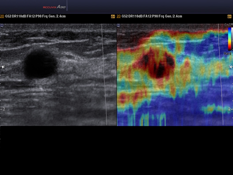
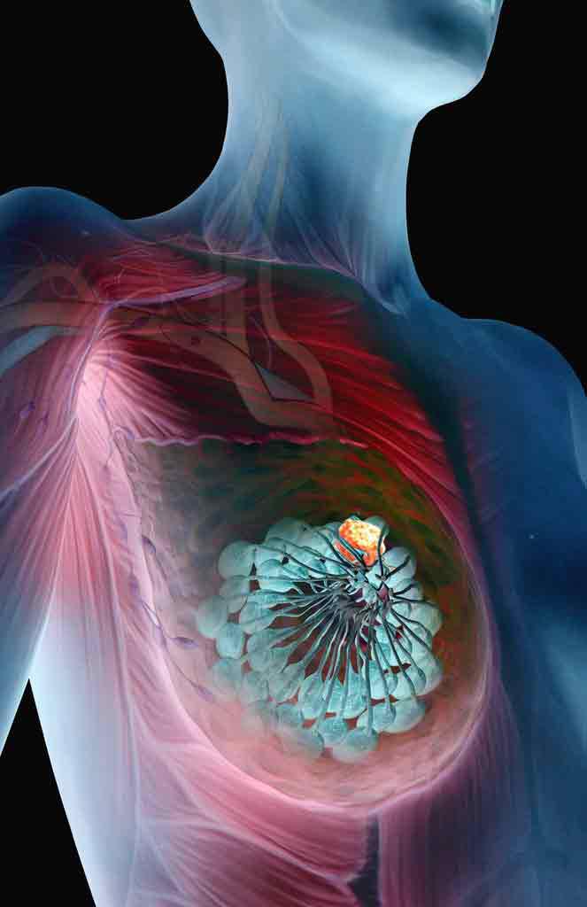
Breast elastography - what is it? During the study, the ultrasonic wave spreads over the entire area of the breast tissue. By means of a specialized sensor, the received information is changed to color images on the monitor. Most often, areas painted in a bright blue color indicate the presence of a malignant tumor in the human body.
Tumor Elastography
Breast elastography - what is it? Breast cancer is considered the most dangerous of the most common diseases in women. Every year, approximately 1 million women identify it.
The main danger of this condition is the absence of symptoms at the first stage of the development of pathology, when it responds well to treatment. If a woman has symptoms indicating a possible presence of breast cancer (severe fatigue, loss of appetite, the presence of pain), then even an urgent surgery does not help in all cases.
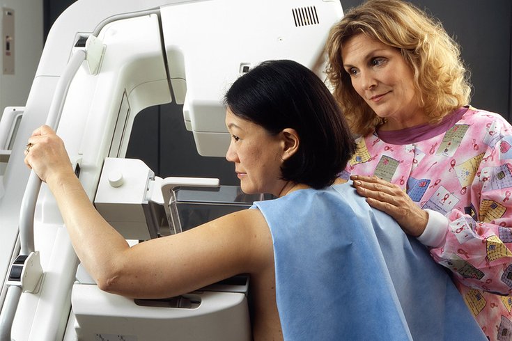
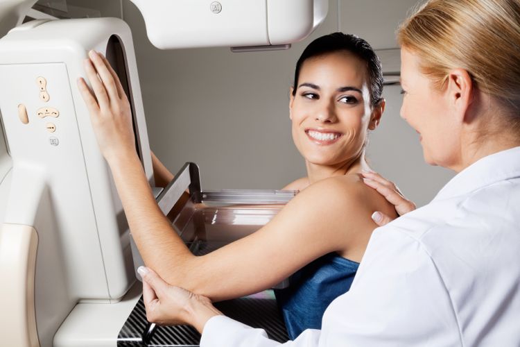
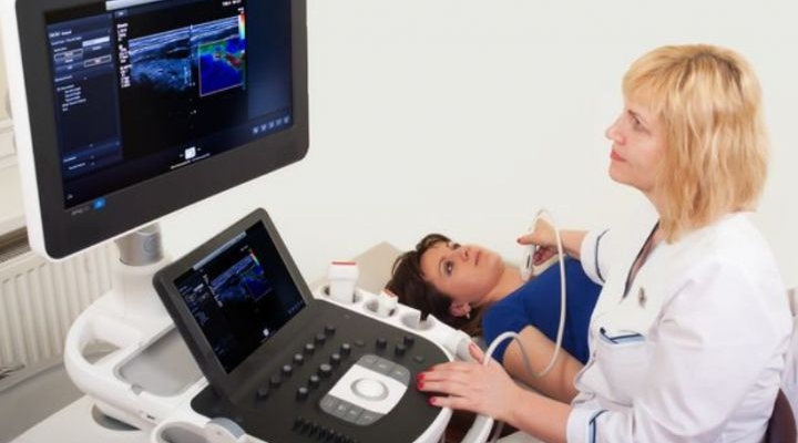
Doctors advise every woman over the age of 25 to undergo an annual ultrasound examination of the breast, and women over forty should undergo a mammogram. At present, elastography can easily compete with both ultrasound and mammograms, and even surpasses them in terms of effectiveness and image quality.
Pros of the procedure
The main advantages of such a survey include:
- identification of benign and malignant tumors in the mammary gland;
- when determining the presence of a benign tumor, a biopsy is no longer necessary;
- the results of the study can be obtained even at the time of diagnosis of the neoplasm.
A poorly distinguishable picture of a malignant formation in the chest at the stage of absence of metastases, vague results after ultrasound and mammography lead to the need to conduct mandatory collection of material for cytological or histological analysis. To take the material into the laboratory, it is necessary to invasively violate the integrity of the tissues by piercing them. Elastography makes it possible to identify the nature of the tumor with a diameter of at least 20 millimeters.
The main distinguishing feature of this research method is the absence of contraindications. Even a woman who is nursing and bearing a baby can be diagnosed without much risk. Elastography provides more accurate information about the tumor and its location.
Diagnosis of ultrasound and elastography
Ultrasound and breast elastography - what is it? When conducting diagnostics and determining the etiology of the neoplasm by means of elastography, the obtained color images begin to be examined in detail by doctors, which allows them to decide on further treatment. For example, in the subcutaneous fatty tissue of the breast, over time, a seal from the connective tissue may occur. As a rule, this is a simple lipoma of a benign nature, although when conducting a simple ultrasound, a large wen on the monitor looks like an extensive neoplasm.
After its discovery, the doctor prescribes the woman an additional delivery of laboratory and biochemical tests, as well as taking a puncture for histological analysis. When elastographic diagnosis on the monitor, a large lipoma is a separate area, painted in green.
This is exactly what neoplasms look like, which are benign, have elastic tissues and do not pose any danger to the patient's health. A biopsy and other examinations that require a lot of time are not necessary.
Reviews about ultrasound with elastography of the mammary glands are mostly positive. In addition to its speed, such a procedure is also useful in that it does not disturb the patient’s peace of mind, does not bring her any stress and allows her to calmly go through the study and wait for the results.
Pros of using the procedure
The advantages of ultrasound elastography of mammary glands with lymphatic surfaces are:
- it is possible to determine the degree of deformation of tissues and the mammary gland as a whole;
- if a malignant formation is detected, the doctor will not have to prescribe additional measures to identify the surgical site - the neoplasm is visualized on the screen without errors;
- does not require preparation of the patient during an urgent examination
- the procedure lasts no more than 30 minutes even when a large number of cysts and tumors are detected;
- the image with good visibility is provided to the doctor;
- high-precision images with the involvement of a doctor of a narrower specialization;
- the procedure does not cause the patient any pain or inconvenience;
- it is possible to immediately establish or refute the presence of a cancerous tumor in the mammary glands; if a woman has symptoms of a malignant tumor, you don’t have to spend time on additional tests, but you can immediately start treatment.

Preventive examination
When conducting a study as a preventive measure through elastography in the breast tissue, the doctor can determine the presence of cellular elements with an admixture of lymph or blood. Such a process indicates the presence of:
- inflammation
- malignant or benign tumors.
Diagnosis of a cancerous tumor helps to immediately begin the passage of drug therapy in order to stop the negative process. The main distinguishing feature of such a diagnosis is the ability to identify a cyst and fibroadenomas using a color picture. To prescribe treatment, you only need to consult a gynecologist.
In some female diseases of the reproductive system, it becomes necessary to assess the condition of the lymphatic vessels and nodes in the chest. In this case, doctors prescribe elastography.
Features of the procedure
Elastography is performed to detect a benign or malignant formation and its elasticity using modern computer technology. When the sensor presses on the examined site of the mammary glands, the adipose tissue quickly deforms, and then restores the initial state, which makes it possible to accurately distinguish it from malignant formation.
The tissues of the cancerous tumor have a high density, therefore, when pressed by the sensor on the monitor of the device, it is well determined that the seal remains within its boundaries or only slightly changes its shape.
By type, elastography is divided into:
- shear wave elastography;
- mammary compression elastography.
Compression elastography
The doctor may prescribe one or another study, depending on the clinical picture. Type 5 breast elastography will be slightly different from the second. When performing compression elastography, the breasts are acted upon by pressing, and the result of the tissue structure and its deformation is shown in full detail on the monitor as a color picture. Further, the result is calculated based on the stiffness of the formation and comparison of the density of fatty tissues on adjacent breast tissues. When conducting compression elastography for compression, it is not the doctor’s hands that are used, but only a readout sensor.
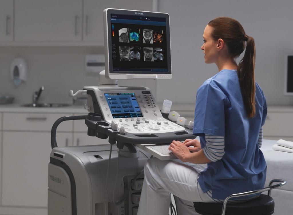
Which device is used?
Recently used devices can do without a sensor, since their sensitivity is especially high. They independently read information about tissue deformation that occurs at the time of the patient's inhalation and exhalation. The tissue elasticity index is coded into a mapping using color and superimposed on the image. Most often, green predominates: this shade reflects fatty, connective and in some cases fibrous tissues. With blue, a biopsy is required.
Shear wave elastography
During shear wave elastography, it is not necessary to press the sensor onto the chest - the device independently shifts the tissues, which guarantees a reliable result. Information obtained from this method of examination is based on the following physical criteria: maximum stiffness, speed of propagation of a sound wave, average statistical data for comparison. The fabric is not deformed under the influence of mechanical compression, an ultrasonic pulse is used for this.
When conducting research using any of the methods described, you can accurately understand if there is education, what is its nature and place of its distribution. Reviews about breast elastography are basically positive. Regardless of the size and depth of the neoplasm, it can be determined in just a few minutes.
The main advantage of such a procedure is the ability to identify the quality of education - a malignant tumor needs to be removed immediately, and a benign tumor must be carefully monitored, and in some cases cured.