The structure of the mammary gland consists of three main parts - lobes, milk ducts and adipose tissue. If the female breast has undergone any disease, then first they carry out diagnostic measures, the basis of which is ultrasound of the organ, mammography and MRI. But despite the good informativeness of the methods, sometimes additional research is required. For example, ductography studying ducts.
What is breast ductography, how this study is conducted, what contraindications the technique has, and much more will be described in the information provided.
The main features of the method
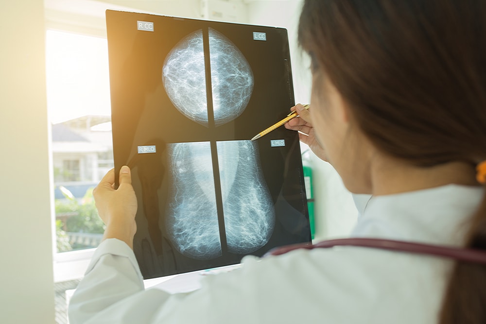
Breast ductography is an X-ray examination of the mammary ducts of the mammary glands. In this case, a contrast agent is introduced into them. Doctors call this method of diagnosis one of the varieties of mammography. Another name for this manipulation, which is often used by medical professionals, is galactography.
What can reveal?
The procedure allows you to identify such violations:
- The narrowing or expansion of the milk ducts.
- Neoplasms, for example, intraductal papillomas or cancerous tumors.
- Pathological areas in the gland.
- Identify their specific location, number and size of formations.
Thus, the main advantage of ductography is the ability to detect neoplasms in the milk ducts, which is not possible for any other method. This is especially important when appointing an operation, since it is possible to identify the exact location of the tumor.
Ductography can also confirm or refute such an unpleasant diagnosis as intraductal papillomatosis. An analysis of the contents of the nipples can also reveal this disease, but it is galactography that puts the final point. It is very important to identify this disease, since it is often a background ailment and a harbinger of oncology.
A referral to the study can be obtained from a mammologist, oncologist, radiologist after ultrasound and MRI of the mammary gland.
As you know, the most dangerous diseases of the mammary glands are cancers. They do not manifest themselves for a long time. Galactography, along with other research methods, allows you to identify the disease in its early stages.
You will read the pros and cons of breast ductography below.
Indications
The study can be shown to women who began to discharge from the nipples of red, brown, and sometimes serous. Doctors will offer to undergo galactography, if there are suspicions of a number of diseases. Indications for breast ductography:
- cancers within the ducts;
- thoracic adenoma;
- papilloma, which is located in the intraductal region;
- diffuse nodular mastopathy or suspected cystic formation.
But you should not rely only on ductography. It is imperative to pass a smear from the nipples for cytological analysis, to detect prolactin levels and breast tumor markers.
Contraindications

The presented procedure has a number of contraindications, in which the study is not prescribed.
Who is contraindicated for breast ductography:
- During pregnancy and breastfeeding.
- Since iodine is part of the contrast medium, the procedure is contraindicated for those who are allergic to this component.
- If the smear showed the presence of atypical cells, then ductography is best not to be performed, since it contributes to their further spread to other tissues.
- With purulent discharge from the papillary region.
- If there are cicatricial changes in the nipple.
- The presence of acute inflammation in the chest, such as an abscess or mastitis.
- Ductography of the mammary gland is contraindicated if a tumor is present in the excretory ducts, and it is palpable. Galactography is contraindicated, since the procedure can contribute to the spread of the tumor further along the ducts.
If there are discharge from the nipples of a different nature, then first of all it will be necessary to pass a smear for examination, and only after the results the doctor will determine the need for ductography.
Preparation for manipulation
Special preparation for ductography is not required. Sometimes the doctor recommends taking antispasmodics, such as Baralgin or Papaverine, a few days before the procedure.
Before research it is forbidden to touch the breast, try to squeeze out the contents of the nipples or massage the mammary gland. Otherwise, there is a risk of injuring the organ.
Many women are interested in whether it is painful to do a breast ductography or not. Do not worry that pain will be felt. Often women experience little discomfort while inserting a cannula into the nipple. It will last just a few seconds. But if the patient is too sensitive, then the doctor can perform anesthetic manipulations.
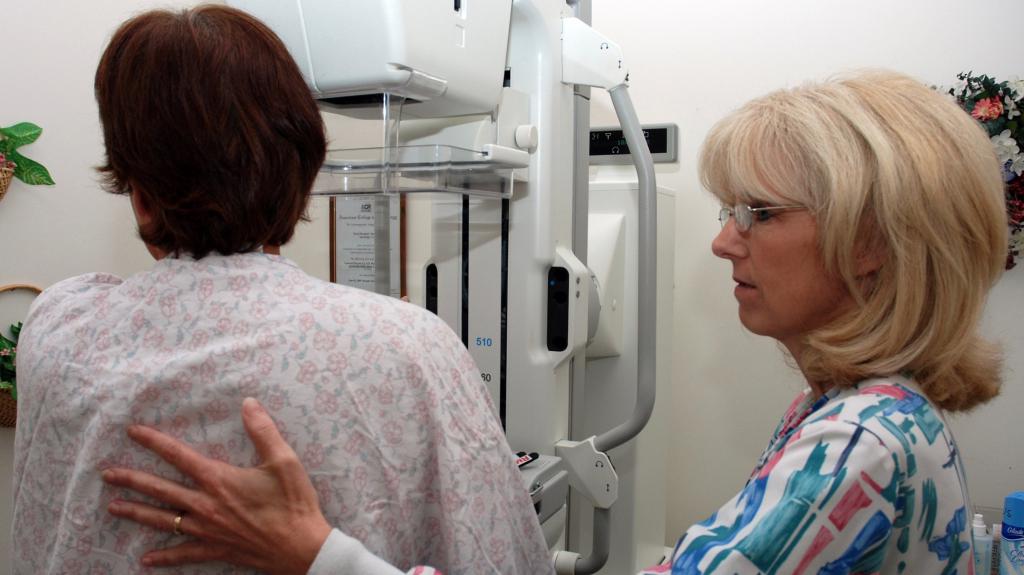
How is the study conducted?
Breast ductography is a method that does not cause much discomfort, only too sensitive women report soreness during the study.
The procedure will be carried out in a separate office, where the patient will be asked to completely undress to the waist, as well as to remove the metal underwear. In this case, the woman takes a horizontal position, namely lies on her side, her hands usually rise up. It is permissible to conduct a study while sitting. But it should be good lighting.
First, antiseptic agents are used, only after that you can proceed to the main manipulations.
At the request of the woman, an anesthetic is initially introduced into the nipple. After the onset of its action, a catheter is inserted into the milky duct. Through it, contrast medium enters the chest. The size of the needle must comply with the following standards:
- Length - 6-8 cm.
- The lumen diameter is 1.0 mm.
Sometimes the doctor advises on the eve of the procedure to take the No-Shpy pill. This is done so that vascular spasm does not occur, since in such circumstances, further actions by the doctor will be useless. Vascular spasm can occur due to the patient’s fear, as a result of a woman’s soreness or nervous state.
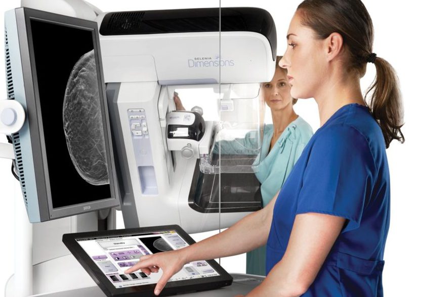
After all the above manipulations, the mammary gland is placed on a special stand of the device. A plate is placed on top of the chest, its pressure allows the contrast medium to completely disperse along the ducts. After that, x-rays are taken in two projections. Then the contrast medium must be removed from the ducts. The entire procedure takes approximately 30 minutes.
As soon as the pictures are transferred to the attending physician, he will be able to decrypt them. If pathological changes are present, additional diagnostics in the form of MRI may be necessary. Only after the final diagnosis is made, the doctor will choose the appropriate treatment.
Disadvantages of the method
Breast ductography is a method that has its drawbacks, one of which is trauma to the milk ducts. This happens due to the introduction of a catheter into them. But this condition does not require special therapy, often such injuries go away on their own.
Possible adverse reactions
It should be remembered that ductography is not a completely safe procedure, so first of all check with your doctor for a list of contraindications. Reviews of breast ductography are diverse, some patients report adverse reactions, others tolerate the procedure easily and painlessly.
As side effects, an allergy to the contrast agent introduced into the chest cavity may develop. Therefore, the doctor first conducts an allergic reaction test. If an incompatibility is detected, then another substance is selected or the research method is replaced by another.
Some patients, on the contrary, note an improvement after galactography and the use of a contrast agent, as the inflammation in the chest passes and the discharge from the nipples disappears.
What is the difference between ductography and mammography?

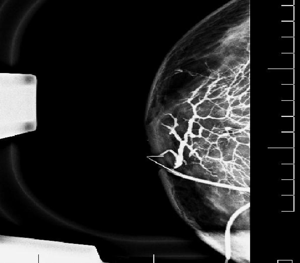
Ductography is a type of mammography. The difference from mammography is that the ducts of the mammary glands are examined by introducing a contrast medium into them. Thanks to this, the doctor can discern well-defined ducts and possible problems associated with it in the pictures.
Only this study is able to identify a tumor process that originates in the ducts of the breast. And also clearly indicates the location of the tumor, which is extremely important during surgery.
In recent years, a disease such as ductal carcinoma is becoming more common. It develops precisely in the ducts of the mammary gland and spreads to other lobes of the breast. Therefore, ductography is considered the most relevant method for detecting the disease at an early stage. Often it is prescribed as an oncology prevention.
Deciphering the study
Breast ductography is a technique by which you can identify the following pathological conditions in the organ:
- Wrong direction and course of the duct.
- The degree of prevalence of the lesion.
- The presence of formations in the mammary gland and their connection with the ducts.
- The presence of enlarged or narrowed sections in the ducts, as well as cliffs.
- The presence of defects and the presence of microcalcifications.
Cancers are indicated by such indicators:
- the appearance of a filling defect - in 14.2%;
- fuzzy boundaries at the entities and destructive areas - at 57.1%;
- in 14.2% of cases - a complete violation of the patency of the ducts and uneven breaks in them;
- the ducts are enlarged, the contours are uneven, the presence of scattered microcalcifications, the presence of cystic enlargements, which is observed in 14.2%.
As soon as the pictures are in the hands of the attending physician, he will be able to decrypt. If he finds pathological changes in the results of the analysis, then an additional examination will be needed to confirm the diagnosis. Only after a few analyzes and instrumental studies can a final diagnosis be made. Then appropriate treatment will be prescribed.