Spiky and sharp, wherever you look: the mechanism of self-sharpening of the teeth of sea urchins
Talking about teeth in people is most often associated with caries, braces and sadists in white coats who just dream of making themselves beads from your teeth. But the jokes aside, because without dentists and established rules of hygiene behind the oral cavity, you and I would only eat crushed potatoes and soup through a tube. And everything is to blame for the evolution, which gave us far from the most durable teeth, which still do not regenerate, which probably pleases the representatives of the dental industry indescribably. If we talk about the teeth of wildlife, then immediately recall the majestic lions, bloodthirsty sharks and extremely positive hyenas. However, despite the power and strength of their jaws, their teeth are not as amazing as the teeth of sea urchins. Yes, this lump of needles under water, stepping on which you can spoil yourself a good part of the vacation, has quite good teeth. Of course, there are not many of them, only five, but they are unique in their own way and able to hone themselves. How have scientists identified such a feature, how exactly does this process proceed, and how can it help people? We learn about this from the report of the research group. Go.
Study basis
First of all, it is worth getting to know the main character of the study - Strongylocentrotus fragilis, speaking human language, with a pink sea urchin. This species of sea urchins is not very different from its other counterparts, with the exception of a more flattened pole shape and a glamorous color. They inhabit quite deeply (from 100 m to 1 km), and they grow up to 10 cm in diameter.
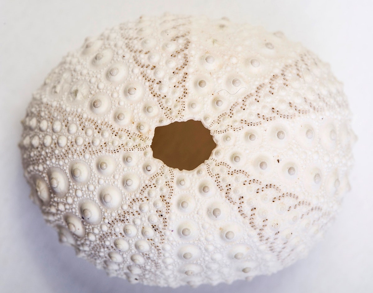
The "skeleton" of a sea urchin, according to which five-beam symmetry is visible.
Sea urchins are, no matter how rude it may sound, right and wrong. The former have an almost perfectly round body shape with pronounced five-beam symmetry, while the latter are more asymmetric.
The first thing that catches your eye when you see a sea urchin is its needles that cover the entire body. Different types of needles can be from 2 mm up to 30 cm. In addition to needles, the body has spheridia (balance organs) and pedicellaria (processes, reminiscent of forceps).
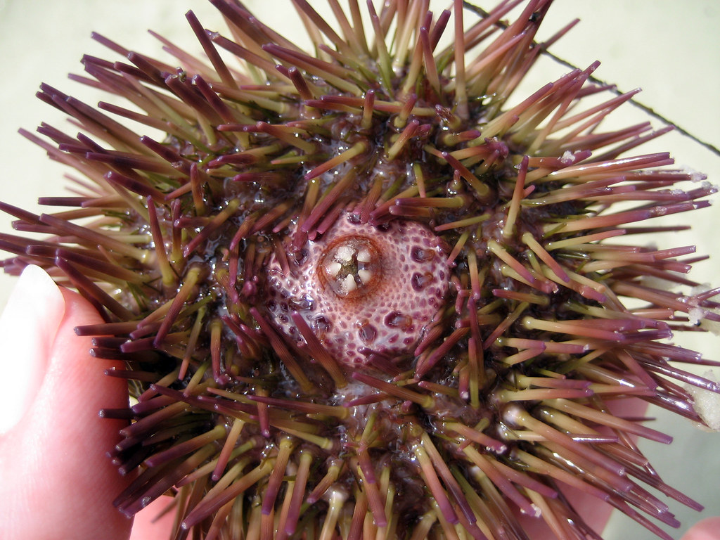
All five teeth are clearly visible in the center.
To portray a sea urchin, you first need to turn upside down, since the mouth opening is located on the lower part of the body, but the other holes are on the top. The mouth of the sea urchin is equipped with a chewing apparatus with the beautiful scientific name “Aristotle's Lantern” (it was Aristotle who first described this organ and compared it in shape with an antique portable lamp). This organ is equipped with five jaws, each of which ends with a sharp tooth (the Aristotelian lamp of the pink hedgehog under investigation is shown in Figure 1C below).
There is an assumption that the durability of the teeth of sea urchins is ensured by their constant sharpening, which occurs through the gradual destruction of the mineralized tooth plates to maintain the sharpness of the distal surface.
But how exactly does this process proceed, which teeth should be sharpened and which not, and how is this important decision made? Scientists tried to find answers to these questions.
Research results
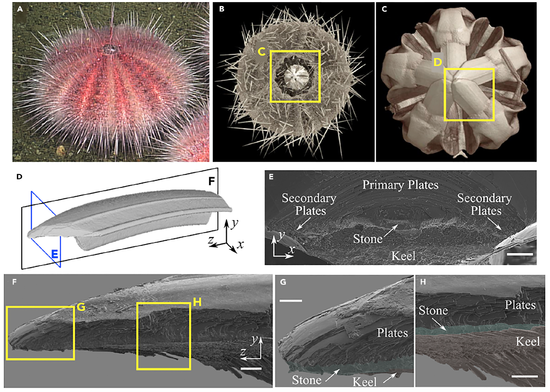
Image No. 1
Before revealing the dental secrets of sea urchins, consider the structure of their teeth as a whole.
Pictures 1A - 1C show the hero of the study, the pink sea urchin. Like other sea urchins, representatives of this species get their mineral components from sea water. Among skeletal elements, teeth are highly mineralized (99%) with magnesium enriched calcite.
As we discussed earlier, hedgehogs use their teeth for scraping food. But besides this, they use their teeth to dig holes in which they hide from predators or it is bad weather. Given such an unusual use for teeth, the latter should be extremely durable and sharp.
Figure 1D shows microcomputer tomography of a segment of a whole tooth, making it clear that the tooth is formed along an elliptical curve with a T-shaped cross section.
The cross section of the tooth ( 1E ) shows that the tooth consists of three structural areas: primary plates, stone area and secondary plates. The stone area consists of fibers of small diameter surrounded by an organic shell. The fibers are enclosed in a polycrystalline matrix composed of particles of calcite rich in magnesium. The diameter of these particles is about 10-20 nm. Researchers note that the concentration of magnesium is heterogeneous throughout the tooth and increases closer to its end, which ensures its increased wear resistance and hardness.
A longitudinal section ( 1F ) of the stony area of the tooth shows the destruction of the fibers, as well as the separation that occurs due to delamination at the interface between the fibers and the organic shell.
Primary plates usually consist of calcite single crystals and are located on the convex surface of the tooth, while secondary plates fill the concave surface.
In image 1G, you can see an array of curved primary plates lying parallel to each other. The image also shows fibers and a polycrystalline matrix filling the space between the plates. The keel ( 1H ) forms the base of the transverse T-section and increases the stiffness of the tooth during bending.
Since we know the structure of the tooth of the pink sea urchin, we now need to find out the mechanical properties of its components. For this, compression tests were carried out using a scanning electron microscope and the nanoindentation method * . Nanomechanical tests involved samples cut along the longitudinal and transverse orientations of the tooth.
Nanoindentation * - verification of a material by pressing a special tool, an indenter, into the surface of a sample.Analysis of the data showed that the average Young's modulus (E) and hardness (H) at the tip of the tooth in the longitudinal and transverse directions are: E L = 77.3 ± 4.8 GPa, H L = 4.3 ± 0.5 GPa (longitudinal) and E T = 70.2 ± 7.2 GPa, H T = 3.8 ± 0.6 GPa (transverse).
Young's modulus * is a physical quantity that describes the ability of a material to resist tension and compression.In addition, in the longitudinal direction, grooves were made with cyclic additional loading to create a model of visco-plastic damage for the stone area. 2A shows a load-displacement curve.
Hardness * - the property of a material to resist the introduction of a harder body (indenter).
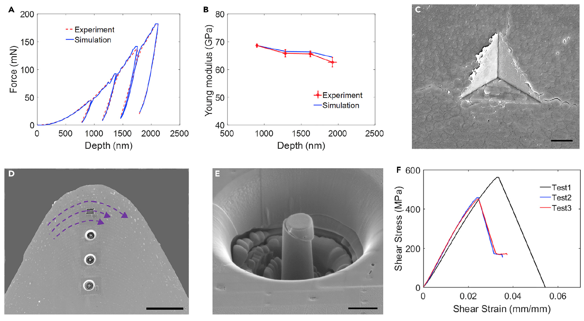
Image No. 2
The module for each cycle was calculated based on the Oliver-Farr method using unloading data. Indentation cycles showed a monotonous decrease in module with an increase in indentation depth ( 2B ). This deterioration in stiffness is due to the accumulation of damage ( 2C ) as a result of irreversible deformation. It is noteworthy that the third development occurs around the fibers, and not through them.
The mechanical properties of tooth components were also evaluated using quasistatic compression of micro-pillars. For the manufacture of micrometer-sized pillars, a focused ion beam was used. To assess the strength of the connection between the primary plates on the convex side of the tooth, micro columns were made with an inclined orientation relative to the normal interface between the plates ( 2D ). Figure 2E shows a micro pillar with a tilted interface. And graph 2F shows the results of shear stress measurements.
Scientists note a curious fact - the measured modulus of elasticity is almost half that of indentation tests. Such a mismatch between the indentation and compression tests is also noted for tooth enamel. Currently, there are several theories that explain this discrepancy (from environmental influences during testing to sample contamination), but there is no clear answer to the question why there is a discrepancy.
The next step in the study of the teeth of a sea urchin was wear tests performed using a scanning electron microscope. The tooth was glued to a special holder and pressed against a substrate of ultrananocrystalline diamond ( 3A ).
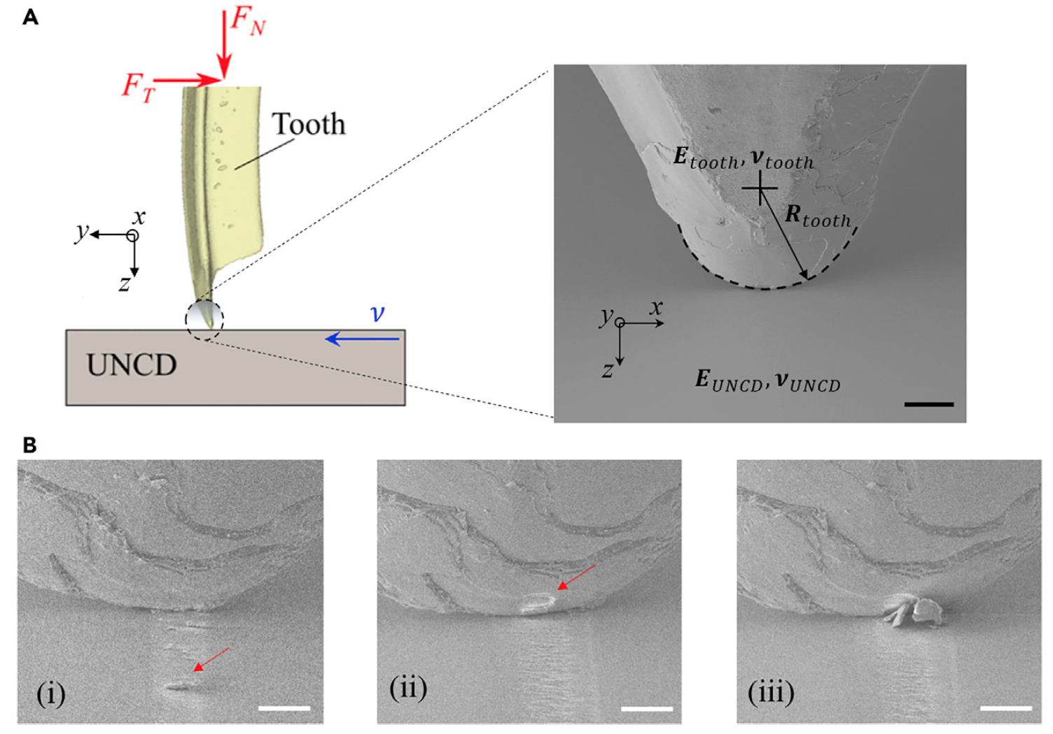
Image No. 3
Scientists note that their version of the wear test is the opposite of what is usually done when a diamond tip is pressed into a substrate of the test material. Changes in the methodology for the wear test allow a better study of the properties of microstructures and tooth components.
As we can see in the pictures, when a critical load is reached, chips begin to form. It is worth considering that the strength of the “bite” of the Aristotelian lantern in sea urchins varies depending on the species from 1 to 50 Newtons. In the test, a force was applied from hundreds of micronewtons to 1 newton, i.e. from 1 to 5 Newtons for the entire Aristotelian lantern (since there are five teeth).
In picture 3B (i) , small particles are visible (red arrow) formed as a result of wear of the stone area. As the area of the stone wears out and contracts, cracks at the interfaces between the plates can occur and propagate due to the compression-shear load and the accumulation of stresses in the area of the calcite plates. Pictures 3B (ii) and 3B (iii) show the places where the fragments broke off.
For comparison, two types of wear experiments were carried out: with a constant load corresponding to the yield strength (WCL), and with a constant load corresponding to the yield strength (WCS). As a result, two options for tooth wear were obtained.
Video of wear tests:
Stage I
Stage II
Stage III
Stage IV
Stage I
Stage II
Stage III
Stage IV
In the case of constant loading, a compression of the region was observed in the WCL test, but no chips or other damage to the plates were noted ( 4A ). But in the WCS test, when the normal force was increased to maintain a constant contact voltage constant, chips and loss of plates ( 4V ) were observed.
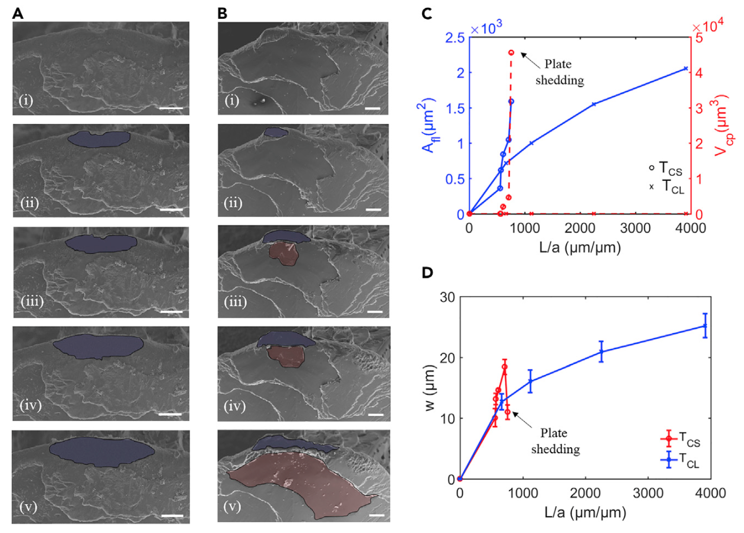
Image No. 4
These observations are confirmed by the graph ( 4C ) of measurements of the compression area and the volume of chipped plates, depending on the length of the slip (diamond sample during the test).
This graph also shows that in the case of WCL, chips do not form even if the sliding distance is greater than in the case of WCS. Inspection of compressed and chipped plates at 4V allows you to better understand the mechanism of self-sharpening of the teeth of a sea urchin.
The area of the compressed area of the stone increases as the plate breaks off, which leads to the removal of part of the compressed area [4B (iii-v)] . Microstructural features, such as the bond between stone and slabs, facilitate this process. Microscopy showed that the fibers in the stone area bend and penetrate through the layers of plates in the convex part of the tooth.
On the graph 4C, a jump in the volume of the chipped area is visible when a new plate is disconnected from the tooth. It is curious that at the same moment there is a sharp decrease in the width of the flattened region ( 4D ), which indicates the process of self-sharpening.
Simply put, these experiments showed that while maintaining a constant normal (not critical) load during wear tests, the tip blunts while the tooth remains sharp. It turns out that the teeth of the hedgehogs are sharpened during use, if the load does not exceed the critical, otherwise damage (chips) may form, and not sharpening.
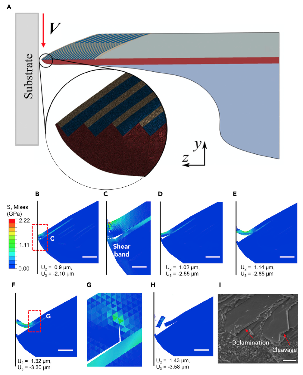
Image No. 5
To understand the role of tooth microstructures, their properties and their contribution to the self-sharpening mechanism, a nonlinear analysis of the wear process by the finite element method ( 5A ) was performed. To do this, we used images of a longitudinal section of the tooth tip, which served as the basis for a two-dimensional model consisting of stone, plates, keel and interfaces between the plates and the stone.
Images 5B - 5H are contour plots of the Mises criterion (plasticity criterion) at the edge of the stone and slab area. When the tooth is compressed, the stone undergoes large viscoplastic deformations, accumulates damage and contracts (“flattened”) ( 5B and 5C ). Further compression causes a shear band in the stone, where most of the plastic deformation and damage accumulate, tearing off part of the stone, leading to its direct contact with the substrate ( 5D ). Such fragmentation of stone in this model corresponds to experimental observations (chipped fragments on 3B (i) ). Compression also leads to delamination between the plates, since the interface elements are subjected to a mixed load, which leads to decohesion (delamination). As the contact area increases, contact stresses increase, causing the crack to nucleate and propagate at the interface ( 5B - 5E ). Loss of adhesion between the plates enhances the bend at which the outer plate is detached.
Scratching aggravates damage to the interface, which leads to plate removal when the plates (a) are split (where cracks deviate from the interface and penetrate the plate, 5G ). As the process continues, fragments of the plate detach from the tip of the tooth ( 5H ).
It is curious that the simulation very accurately predicts chipping both in the area of the stone and in the area of the plates, which scientists have already noticed during observations ( 3B and 5I ).
For a more detailed acquaintance with the nuances of the study, I recommend that you look into the report of scientists and additional materials to it.
Epilogue
This work once again confirmed that evolution was not very supportive of human teeth. Seriously, in their study, scientists were able to examine and explain in detail the mechanism of self-sharpening of the teeth of sea urchins, which is based on the unusual structure of the tooth and the correct load on it. The plates covering the hedgehog's tooth exfoliate at a certain load, which allows you to keep the tooth sharp. But this does not mean that sea urchins can crush stones, because when critical loads are reached, cracks and chips form on the teeth. It turns out that the principle "there is power, no mind," certainly would not bring any benefit.
You might think that researching the teeth of the inhabitants of the deep sea does not bring any benefit to humans, besides satisfying insatiable human curiosity. However, the knowledge gained during this study can serve as the basis for the creation of new types of materials that will have properties similar to the teeth of hedgehogs - wear resistance, self-sharpening at the material level without external assistance and durability.
Be that as it may, nature has many secrets that we still have to reveal. Will they be helpful? Maybe yes, maybe not. But sometimes even in the most complex studies, sometimes not the destination, but the journey itself is important.
Friday off-top:
Underwater forests of giant algae serve as a gathering place for sea urchins and other unusual inhabitants of the oceans. (BBC Earth, voice-over - David Attenborough).
Thank you for your attention, stay curious and have a great weekend everyone, guys! :)
Underwater forests of giant algae serve as a gathering place for sea urchins and other unusual inhabitants of the oceans. (BBC Earth, voice-over - David Attenborough).
Thank you for your attention, stay curious and have a great weekend everyone, guys! :)
Thank you for staying with us. Do you like our articles? Want to see more interesting materials? Support us by placing an order or recommending it to your friends, a 30% discount for Habr users on a unique analog of entry-level servers that we invented for you: The whole truth about VPS (KVM) E5-2650 v4 (6 Cores) 10GB DDR4 240GB SSD 1Gbps from $ 20 or how to divide the server? (options are available with RAID1 and RAID10, up to 24 cores and up to 40GB DDR4).
Dell R730xd 2 times cheaper? Only we have 2 x Intel TetraDeca-Core Xeon 2x E5-2697v3 2.6GHz 14C 64GB DDR4 4x960GB SSD 1Gbps 100 TV from $ 199 in the Netherlands! Dell R420 - 2x E5-2430 2.2Ghz 6C 128GB DDR3 2x960GB SSD 1Gbps 100TB - from $ 99! Read about How to Build Infrastructure Bldg. class c using Dell R730xd E5-2650 v4 servers costing 9,000 euros for a penny?
All Articles