Many women in the mammary glands may have seals while they are expecting a baby. It is during these months that prolactin, estrogen and progesterone are produced in the body, and in very large quantities. As a result, large seals can be felt inside the chest. Some time after birth, the level of hormones decreases, and everything goes away. Formations on the skin of the chest are different, as well as inside it. About them - further.
Causes
Very often , chest pains and formations are noticed by mothers who breastfeed the baby. This is due to clogging of the milk duct or when an inflammatory process occurs. This problem in the medical field is called mastitis. If this diagnosis is established, then the size of the formation can reach quite large, since infiltration always accumulates around the inflammation.

Of course, seals appear not only during lactation, but also when:
- mastopathy;
- cyst;
- thrombophlebitis;
- neoplasms of various etiologies.
Formations may occur inside the mammary glands due to injury or when using not very comfortable underwear. The malfunction of the thyroid gland and the organs that are responsible for reproduction also negatively affect the condition of the breast. An abortion, menopause at an early age and constant stressful situations often provoke the development of tightness in the chest.

Symptoms
Cancer itself is a very insidious disease that is difficult to identify in the early stages. It practically does not manifest itself and does not manifest itself, so it is very difficult to determine the first signs of a tumor in the chest. But in case one of the symptoms was noticed, you must immediately go to a specialist for diagnosis.
- With the appearance of a painless, but at the same time dense formation in the chest, it is necessary to sound the alarm.
- In case there has been a change in the shape of the mammary gland.
- If wrinkling or tightening of the skin in the chest area has occurred.
- There was discomfort or pain.
- A swelling on the nipple or a seal is also a signal.
- Red discharge appeared.
- Enlarged lymph node in the armpit.
In no case should you lose sight of all these symptoms, even if the appearance of changes in the body does not cause you discomfort, you should not think that everything will pass without a trace, because, perhaps, this is the first sign of the appearance of cancer, which is difficult to diagnose on early stages. If you suspect, then you must immediately contact a specialist for diagnosis.
Formations inside the mammary gland can be divided into two groups: these are benign and malignant.

Benign formations
If we talk about the benign type of seals, then they are characterized by slow growth and almost complete absence of aggressiveness. They are located in such a way that nearby tissues remain intact. Always appear in healthy tissue and in the future can not cause the development of oncology.
In some cases, such nodules in the chest can significantly increase the risk of developing a malignant tumor.
Types of tumor
In accordance with the classification according to histology, several types of breast tumors are distinguished.
- Fibrous formation in the chest has a glandular origin, that is, it is based on connective tissue. Usually, only one ball becomes noticeable, and its size sometimes reaches 7 centimeters. Some may have several at once. The risk group includes young girls. The main feature of focal formation in the chest is associated with the complete absence of any symptoms. Changes in the chest can be detected only when examined by a specialist.
- A cystic formation in the chest is a cavity enclosed by walls and filled with a liquid component. The appearance is directly related to the obstruction of the ducts of the mammary gland. With a cystic formation in the chest, there are practically no symptoms, so only a specialist will help to identify changes. If we talk about treatment, then first prescribe special medications, and in the future, a puncture may be required.
- A tumor in the chest with a loose consistency is called a lipoma. When probing with fingers, it does not cause pain and does not change its position. It can increase, but it happens extremely slowly. Almost always, a lipoma remains a benign formation. Only in units does it degenerate into liposarcoma. Great risk extends to people over 50. In any case, doctors advise careful monitoring of the seal. If it has significantly increased, then you will have to resort to a radical method, that is, sectoral resection.
- Papilloma is manifested by certain symptoms, so it can be cured already at an early stage of development. In most cases, it affects the skin near the nipple. If you do not start treatment, then after a lapse of time the education goes into oncology. The most pronounced symptom of this disease is associated with the release of bloody fluid, and when pressed, severe pain is felt. Papilloma cannot be cured by conservative methods, so the specialist recommends a sectoral resection. In other words, it is simply cut out using surgical instruments.
- For malignant breast tumors, a high risk is characteristic, since quite often the disease leads to death. Modern medicine does not stand still, and now experts can accurately diagnose oncology even at an early stage. It occurs due to hormonal failure in the female body.
Cancer factors
Breast cancer is a leader worldwide. Based on statistics cited by WHO, every day doctors identify about 1 million cases of the development of just such a disease.
Doctors call a number of factors that trigger the occurrence of this health problem. This may include:
- smoking cigarettes;
- the inability to become pregnant and give birth;
- the presence of excess weight;
- diabetes mellitus of any type;
- menopause starting later in life;
- hereditary predisposition;
- excessive consumption of alcoholic beverages.

Malignant tumors
In medicine, more than 15 varieties of breast cancer have been identified. Most often, it affects the ducts and individual lobes of the chest.
In total, 4 degrees of cancer development are distinguished, and there is also one preliminary.
- A precancerous condition, that is, a seal appears, but it has not yet passed to the organs located nearby. If the doctor revealed a problem, then the patient had a predisposition to the disease.
- Stage 1 is represented by an invasive form, that is, the disease gradually spreads to nearby tissue. Usually the affected cells quickly go beyond the main focus. It is important to carefully monitor your health and not neglect scheduled examinations by a doctor. If cancer can be detected at this stage, appropriate measures will be taken. During this period, the formation can grow up to 2 centimeters.
- At the 2nd stage, the size increases to 5 centimeters and there is a lesion of the lymph nodes located in the armpits. When adhesions with healthy tissues appear, another stage of the course of the disease begins.
- From the 3rd stage, you can visually notice the first symptoms, that is, the mammary gland changes its shape, and red spots appear on the skin.
- At the 4th stage, there is practically no reason to carry out treatment, since during this period most of the organs are already affected by cancer cells.
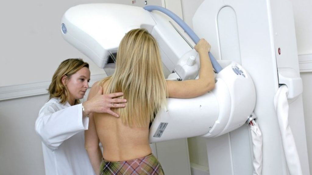
Diagnostics
A seal may appear inside the mammary gland, and this happens to women of any age. If in time to identify changes in the chest and consult a specialist, it will be possible to significantly increase the chances of a complete cure. The specialist should prescribe a full examination, and then choose the appropriate treatment methodology.
In modern medicine, the method for diagnosing chest lumps has been greatly simplified. A fairly large number of devices have now been developed that in a matter of minutes will give an answer to any question.
All patients with education in the mammary gland are sent to such studies as:
- X-ray (young age prevents you from reading the picture correctly, since there is a large volume of glandular tissue in the chest ) ;
- Ultrasound
- galactography, which is a type of mammography (a specialist introduces a drug for contrast x-ray into the milk duct and reveals seals from the inside);
- a biopsy or puncture helps to accurately determine the nature of the formations (the doctor makes a small sampling of the breast tissue and sends it for further research).
It is worth remembering that if the formation in the chest is anechoic, then it is not displayed on ultrasound. It does not lend itself to sound vibrations. Anechogenic formation in the chest is quite difficult to diagnose. But it develops into malignant only in rare cases.
In turn, with hypoechoic formation in the woman’s chest, black spots appear in the picture.
If we talk about seals in the chest, then this is not always an oncological disease. In many cases, this symptomatology indicates a cyst or fibrocystic disease.

Treatment
It is worth recognizing the fact that self-medication in this matter can have an extremely detrimental effect on the female body. That is why it is necessary to contact a specialist who will conduct an examination, collect an anamnesis, find out the patient’s medical history, get acquainted with the problems of heredity, conduct an examination, find out about internal disorders and only after that plan the treatment complex.
Depending on what characteristic the compaction has, and therapy will be determined. Therefore, in the case of a neoplasm in the area of the mammary gland, self-medication is not recommended, because only a specialist can establish a benign tumor or it carries extremely dangerous consequences.
Lactostasis is one of the types of breast diseases, in which case the doctor recommends various compresses, for example, based on camphor oil or Vishnevsky ointment.
If the diagnosis of "mastitis" was made, in this case it is necessary to take antibiotics, if an abscess has formed, resort to surgical intervention. This disease should never be triggered.
If a woman was diagnosed with "mastopathy", then it will be necessary to remember the fact that you need to visit a doctor every six months in the future in order to monitor the dynamics of the disease, in parallel with this you will need to take a complex of medications.
Doctors also recommend that young girls visit a mammologist once every 2 years so that he will conduct an examination and, if any seals are detected, immediately proceed to diagnosis and treatment. For older women, it is recommended that this be done once a year, as well as an ultrasound scan.
What are the treatment methods?
When compaction in the mammary glands, various hormonal and non-hormonal drugs are prescribed. If the education is diffuse, then therapy will be based on the cause of the pathology. After all, it was this reason that caused the dysfunction of the pituitary gland, and a failure occurred in the ovaries. In this case, treatment begins with reproductive organ therapy. Normalize their work, improve the work of the liver and nervous system. In this case, hormonal drugs, androgens, oral contraceptives are prescribed, they are designed to inhibit the production of mammotropin.
If a diagnosis of "nodal mastopathy" was made, then in this case, drugs intended for immunotherapy are prescribed.
In most cases of the occurrence of mastopathy, surgical treatment is prescribed. In this case, the surgeon is committed to removing nodes. But with nodal mastopathy, the non-hormonal method is also used, it is aimed at vitaminization, non-steroidal anti-inflammatory drugs are taken, as well as drugs that improve blood circulation.
The patient is recommended to use antioxidants, phospholipids, they help reduce the load on the liver. Important at the time of treatment of such a mastopathy is a properly selected bra. In no case should you go to saunas, a solarium, you need to minimize exposure to the sun.
If a cyst of 0.5 mm was detected, then in such cases a very conservative treatment is prescribed, first of all, the hormonal background of a woman is normalized. In order for a single-chamber cyst to resolve, a puncture is necessary. The specialist performs a puncture, pumps out the liquid, introduces a special solution that will destroy the capsule. But if an atypical cyst was detected, then in this case it is necessary to perform an operation and remove the affected tissue, and then send it for histological examination to exclude cancer.
There are frequent cases of using the aspiration method. A special device is introduced into the cavity of the cyst, which is engaged in pumping out the liquid, and if traces of blood are visible in this liquid, then an additional study is prescribed. Drug therapy in this case is hormonal, drugs are also used that can strengthen the immune system.
If you timely detect a disease, densification or swelling and immediately consult a specialist, in many cases it will be possible to cure this without surgical intervention. Timely diagnosis helps to avoid serious consequences. In the early stages, tumors can be cured with the help of pharmacological preparations, medicines based on herbs, ointments and compresses.
Prevention
In order to prevent the disease and not give it any chance to develop, it is necessary to adhere to several principles.
- A regular breast examination is necessary.
- During an independent examination, assess the color of the skin, if there is a change, then you need to go to the doctor.
- Pay attention to the nipples, they should not be retracted, should not differ in color.
- The chest during the absence of menstruation should be evenly soft.
- In the event of any seal in the breast tissue, you must consult a doctor.
- During an independent examination of the mammary gland, palpation must be done both horizontally and vertically.
- In order to conduct a full examination of the entire breast and not miss the slightest seal, the breast must be divided into zones conditionally.
- After the palpation of the breast has been done, it is also necessary to check the armpits, evaluate their size and fix the density of the lymph nodes.
- As soon as any seals and signs that a formation has appeared in the chest have been detected, it is immediately necessary to go to a specialist.