A little about the internal warmth
Medicine is an extremely important area, but, unlike most sciences, for a very long time it was an unsystematic set of practices that, to one degree or another, relied on the culture of society. This, as they say, a medical fact, and nothing can be done.
Traditional medicine flows in the veins. Very many concepts and techniques that are now in full use, were formulated more than two thousand years ago. On the one hand, this is very cool - for so much immense experience has been accumulated, and on the other hand, all new ideas are forced to compete with good old techniques that are familiar to doctors from college days, and therefore do not require efforts to master something new. .
However, over the past 250 years, the challenges facing medicine have become more complicated by orders of magnitude, and this trend has maintained its acceleration, so that new complex technologies cannot be avoided.
Now it's time to introduce yourself. My name is Anna, a few years ago, after graduating from a good school, I broke the template and went to study at the medical faculty. Now I am a surgeon with a tender love for the exact and natural sciences.
It so happened that I got to "drive" a pocket thermal imager, which is connected to the micro usb connector of the phone. And of course, as a raccoon drags everything into the water, I dragged a new toy to work ...
Attention! The article contains photos taken during the operation. All of them are processed and cleaned under spoilers. But if you have weak nerves, then perhaps you should refrain from viewing.
Yes, everything.
I will not discover America by stating that the temperature is normal in different parts of the body. The heat source in our body is energy metabolism reactions — when proteins, fats, and carbohydrates are “converted” into the body's internal energy currency, ATP. All these reactions are exothermic, i.e. occur with the release of heat. The main conductor of heat in the body is blood. With great speed, it moves from deep-lying organs to the surface of the body and back. Accordingly, areas of the body where a well-developed network of capillaries with a relatively large diameter will be warmer than areas of the body where the vessels have narrowed or their network is not so developed. (You must have seen the classic posters of brush temperature before and after smoking a cigarette).
It is also unlikely that anyone will be surprised that areas of the body affected by the disease may have a different temperature than normal - during the inflammatory process, a cascade increase in metabolism and expansion of small vessels occurs.
One of the mastodons of ancient medicine, Avl Cornelius Celsus, formulated the signs that tell about inflammation - redness, swelling, increase in local temperature, pain (Latin rubor, tumor, calor, dolor).
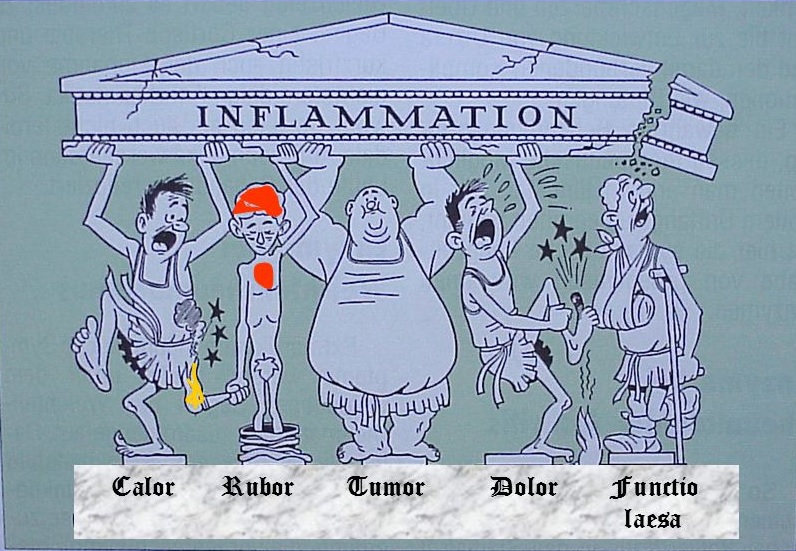
The fifth column of this composition, the “loss of function,” was formulated somewhat later and, for my taste, is not so much a full member of the team, but a logical consequence of the preexisting quartet.
At the same time, tissues that have lost their vitality become colder than normal. This is achieved due to the lack of metabolism and violation of the vascular network.
In fact yes. And now in order.
Medical devices are very expensive to manufacture and maintain. This concerns everything up to alloys for surgical instruments, and it’s not worth talking about high-tech devices. However, as a certain method becomes sufficiently studied and developed, there is a tendency to simplify it and reduce the price. My favorite example is the ability to use a smartphone to display images during laparoscopic operations. The hospital saves a lot of money on the purchase of special equipment for displaying a signal on the screen, color quality settings, and a special lamp. Nobody argues that a smartphone is not an ideal replacement, but thanks to this simplification, minimally invasive surgery has become available in a number of third world countries.
Studies using professional thermal imagers have been conducted for a long time. The analysis of modern publications is given a little lower, and from a long-standing one, which have already become a textbook of the temperature of a person’s hand before and after smoking a cigarette.
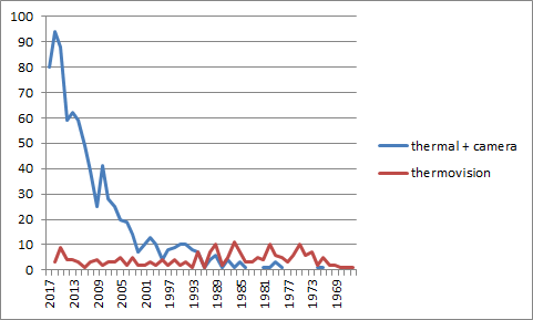
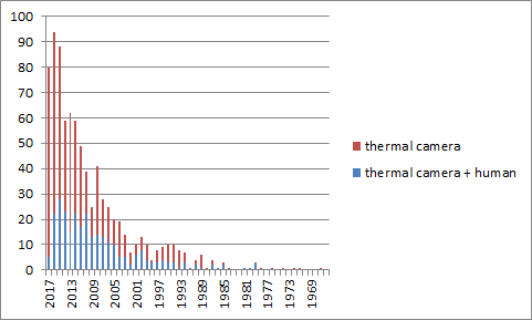
At the same time, as the analysis of PubMed distribution showed, interest in the topic of temperature in a medical-biologically oriented environment appeared about 50 years ago. Judging by the statistics of the issue, up to 1992 articles in the issue at the request of “thermovision”, were no less than articles at the request of “thermal” + “camera”. Perhaps there is still a linguistic effect: the word “camera” 50 years ago was not so popular - but I’m not sure about that at all.
My attempts to appeal to the world mind give an ambiguous result: at the thermovision camera request, PubMed issues only 36 articles, with an amusing distribution by terms and specialties — cosmetology and dentistry prevail among medical fields by a large margin. Also, one group of neurologists from the Czech Republic published three articles on muscle pain, judging by the publication dates, we are talking about the same study, but splitting into three publications increases the proportion of pain studies in the overall picture. Not satisfied with this situation, I asked PubMed to search for “thermal” AND “camera” also in all databases.
In addition, I was curious how often these studies were applied to people.
It turned out that thermal imagers in particular were much more often used in the context of the study of people than methods of visualization of temperature regimes in general.
The issue of “thermal” AND “camera” turned out, of course, with a huge number of false-positive inclusions. I looked through the headlines and abstracts of the 500 most recent publications (total issuance of 830 points). As a result of this work, a lot of things were eliminated that in my subjective view are not applicable to clinical medicine - this list included articles on heat-conducting alloys, a lot of meteorological research, analysis of human recognition algorithms, nuances of electron microscopy protocols, and determination of alcohol content in beverages. I also referred to non-medical articles on the effects of laser acupuncture on the intestinal energy meridians.
Among these five hundred publications were articles for the years 2010 - 2017. I dropped 2010 and 2017 as incomplete and made a schedule of the “popularity” of each of the topics in individual years.
I defined the subject of the article subjectively, but tried to adhere to more or less simple criteria. The topic of Biology includes everything related to the study of wild animals, veterinary medicine, and the meat and dairy industry. To the topic of physiology, I carried mainly experimental studies on one or another biochemical mechanisms in the experimental organism. As you can see from the graph, the main areas of interest are the same.
Here is the integral division by topic in the entire sample:
And here I would be happy for my colleagues - after all, various surgical specialties occupy an impressive part of the resulting “pie”, but I wasn’t sitting in one place again and I decided to check for what purposes temperature measurement is used in each article.
In this analysis, I removed from consideration of a group of articles on biology and physiology, as having no direct relation to clinical medicine, and also refused from dentistry, in which I understand almost nothing.
I marked the articles with the “diagnostics” tag, when temperature mapping indicators were used to identify the patient’s disease, or determine the stage of the disease, the location of the affected tissues in the body. The “treatment control” tag refers to those cases where the thermal sensor was part of the feedback mechanism: in real time, it evaluated the methods of treatment used - for example, it stopped working when the instrument overheated, or marked the treated areas of the skin.
The “treatment control” group mainly included articles on the use of laser in various fields - cosmetology, ophthalmology, neurosurgery, oncology, and many other places. On the methods of diagnosis was emphasized in the articles on diabetic defeat of the feet, various vascular diseases. Ophthalmologists have investigated the ability to diagnose dry eye syndrome. The popularity of epidemiological studies has come down to one question - the in-line measurement of passenger temperature at international airports. It seems to me that the topic is not as scientific as resonance.
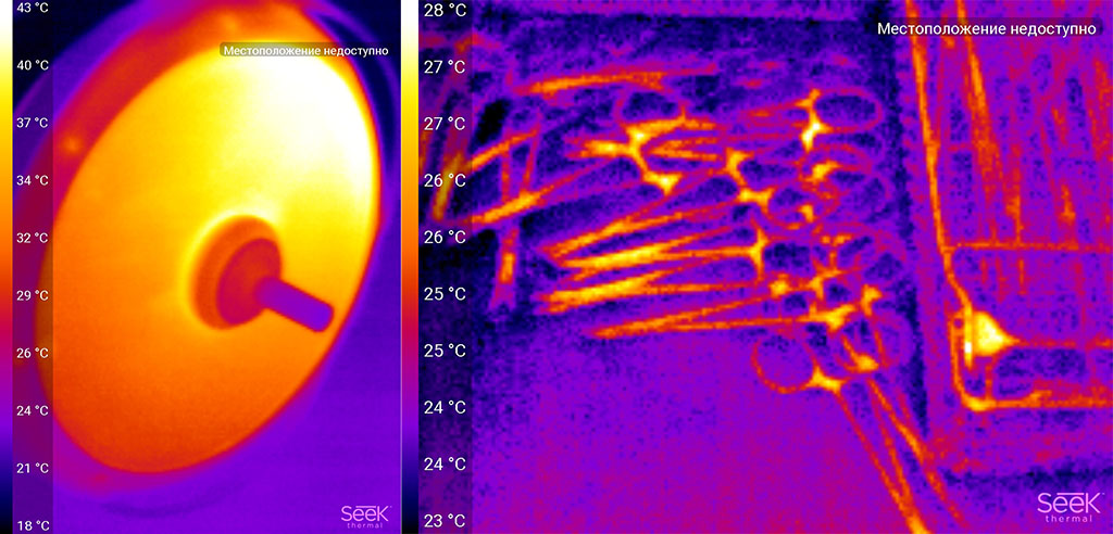
The share of "diagnostic" research in surgery is offensively small. In most cases, the authors tried to focus on the control of heating of surrounding tissues when working with various types of coagulating devices. How hot is it? Well, according to my measurements, something like this:
The black line is the tip of the coagulator, the white dot is the working surface at the very end of the tip + tissue that it touches. As you can see, with an average temperature of the surface of organs 31-35 * C, the coagulator heats up to 55.
And this is what a portion of the greater omentum treated with coagulation looks like (diffuse bleeding was stopped).
Such temperature regimes are consistent with what is written in the articles. Good or bad - no one will say. At a distance from large vessels, working at such temperatures is quite acceptable. Fichey must be properly used so that there are no bugs.
I did not have a clear concept on what to shoot. Moreover, the results of my theoretical studies help only conditionally - in part of the articles they directly write that they used infrared radiation sensors, in part - that they used a professional thermal imager, and sometimes they don’t understand what the chip is all about - it’s only clear that they detected the temperature and made a conclusion from this.
In general, I was looking for what phenomena can visually demonstrate a snapshot of a thermal imager.
Hope to shoot everything and quickly disappeared in large numbers for two reasons: the laws of maintaining sterility are not always possible to get into the operating wound with a telephone, and technical - the technology of interventions is such that on the one hand the organs are in close contact and maintain each other's temperature and, on the other hand, we often use instruments that locally heat fabrics to high temperatures. If the first was clear in advance, then I could not imagine the scale of the second effect.
A healthy loop of the small intestine and mesentery with feeding vessels. The imager traces even thin vessels that are just not visible. In daily practice, these vessels are detected either when looking at the mesentery "for lumen" (badly helps with marked edema, lymph nodes, adhesions), or by touch by pulsation. Brown - surgeon's glove.
And this is the colon, after it was mobilized - freed from the ligaments that fix in the body. It can be seen that the feeding vessels feel very well and the intestinal wall is evenly stained on the thermogram - there is no edema or weakening of the blood flow.
The picture was taken during a stomach operation. The remaining part of the stomach is visible on the right (indicated in green). It can be seen that the entire wall is uniform in temperature signal. The purple stripe (marked with green hatching) corresponds to the gland resection line - there the tissues are “sealed” with a coagulator and in the future they will form a scar.
Blue highlighted clip, superimposed on the duodenum. It can be seen that the tissues above the place of clamping are colder - they do not have blood flow, they will be removed during the reconstructive phase of the operation.
Another operation resection of the stomach. The color indicates the clamp on which the output section of the stomach, cut off from the duodenum. It can be noted, unlike the previous example, that the stomach sections close to the clamp are colder - the fact is that one of the arteries supplying this section is already bandaged.
Beauticians have long appreciated the benefits of temperature mapping for their specialty. As was shown above, more often the task of measuring temperature is the quality control of the procedure performed.
Nevertheless, judging by the results of my research, doctors use thermal imagers or infrared cameras to diagnose various conditions on the skin temperature - mainly neurologists shared this niche with the study of chronic muscular pain and sports medicine doctors who are interested in everything - and overworking muscles, and heat transfer in sports underwear, and, what intrigued me most, was scanning of the subcutaneous tissue with subsequent analysis of its thickness and biochemical properties.
But this is all the lyrics. In our department, it is possible to remove plenty of gradually healing postoperative wounds.
Here is the wound at the time of the end of the operation:
In addition to the factors described, the photograph illustrates the uniformity of ligatures, as well as the place of thinning of the subcutaneous tissue (oblique fold in the upper part of the photo). It exists for a long time due to the specific deformation of the spine of the patient.
I work in a planned surgical hospital without purulent separation. Inflammatory processes with the wounds of our patients are quite rare, and we are working to make it happen even less often. The patient from the next photo as one of the stages of the operation closed the colostomy - the exit of the colon to the anterior abdominal wall, which was formed a few years ago. Since the presence of a colostomy is a prerequisite for the inevitable contact of an operating wound with intestinal microflora, such an operation is called “conditionally infected” and according to statistics in such patients complications occur more often during wound healing.
As I already said, my first attempts to photograph something were a failure. You cannot shoot with a thermal imager according to the same principles as with a camera. When photographing, I usually waited for the local work area to be clearly visible, and took a general picture - it was obvious who was doing what and in what area. With a thermal imager, this number does not pass - the surgeons' hands do not contrast with the patient’s organs.
A typical situation when I asked everyone to remove hands, napkins, tools from the wound and quickly filmed what happened:
In fact, this frame has a rather large surface of the liver (left), the output section of the stomach (highlighted on the right), the small intestine is the right edge of the frame. Everything except the gallbladder (hatched) merges.
The next step was to “hold on the weight so that it was visible”. Organizational problems are already connected with this - not only I (the assistant) are distracted to take off gloves, take pictures, then wash my hands again, put on a new set of dressing gowns and gloves, but the operating surgeon has to postpone work and help me. And still, success is dubious.
But it turned out to be a problem to be solved. It was necessary to somehow neutralize the effect of heating from neighboring organs - then the picture will correspond to the tree of blood vessels. For example, wrap the organs marked for a photo in a napkin moistened with a solution of room temperature. The number of visual frames has increased significantly.
The idea was not new, the dentists had already described the method for determining teeth cutting out in children, when the oral cavity was first blown with cold air, and then the temperature of the gums were recorded.
In my case, the methodology has led to a very clear visualization of a colon tumor.
In the issue I analyzed with PubMed there was one article from the transplant section. In it, the temperature of the surface of the abdomen tried to determine the level of metabolism after bowel transplantation. I suspect that many technical details awaited the researchers on this path, but I’m talking about something else. Transplantation of the intestines is not limited, and the assessment of the adequacy of the blood supply there is very important.
Most often, the blood supply of the graft is estimated by its color - during the preservation period the organ is washed with a colorless solution, therefore the tissues become yellow-brown-gray. And after the start of blood flow stained in the range from pink to red and lilac.
The thermal imager shows that the tissues located under the organ capsule along the resection line are worst of all blood supply, which is quite logical, since during the work there was a thermal damage to the blood capillaries and the tissue itself. The fact that the right segments look much warmer is an artifact, they warmed up from neighboring organs. In this case, given the uniformity of color transition, we can say that the perfusion is adequate.
Sometimes, to cause reflex relaxation of the vascular walls, the graft is easily massaged. The video shows how after that the temperature of the entire surface rose (the tissues warmed up a little by hand), but then the kidney itself retained this temperature due to improved blood flow, and a piece of fat cooled to its original state.
Unfortunately, the revolution “came, photographed, made a diagnosis” did not work.Of course, the majority of routinely used methods (assessment of tissue color, tone, peristalsis) are subjective and appeal to the doctor’s experience, but at the same time, the principle “if you doubt the viability of tissues, consider that it is not viable”. And hardly anyone agrees to preserve fabrics that look dubious outwardly, even with normal indicators of technology.
I would like to share my impressions into several areas:
Ease of use in the operating room is low: first, the device cannot be placed in a sterile casing, therefore either you need to ask someone (who doesn’t understand very quickly what it is), or wash your hands several times, which spends a lot of time, gloves, gowns. Secondly, it is difficult to direct the camera exactly where it is necessary - the beam is narrow and does not come from the place where the camera is usually located. The latter is partly solved by turning the phone over, but not that this is the perfect solution. The most organic solution I personally see is an aquabox-type casing that covers everything except the lens, a Micro-usb extension cable, so that the phone can lie to the side.
The second block of impressions concerns the medical side of the issue. An analysis of the literature showed that the direction of temperature mapping in medicine is promising, but it requires elaboration of the methodology (such as the idea of pre-cooling the subject), and needs an “ecological niche” - a field of application where mapping would open up new possibilities. There are examples of successful solutions - diagnosis of trophic disorders in patients with diabetes. I personally wonder if it is possible to evaluate the functioning of the graft through these methods. If due to such a non-invasive and conditionally cheap procedure, it would be possible to more accurately select therapy in the postoperative period, it would be very cool!
Many thanks to my husbandwho agreed on a thermal imager and edited the article, and colleagues for their responsiveness, patience and assistance in filming.
The SeekThermal thermal imager was provided by the company Dadzhet, the official distributor of SeekThermal in Russia. Dadjet suggested that I publish the promotional code Thermal Imager for the readers of this article, which gives a 10% bonus on its purchase.
Traditional medicine flows in the veins. Very many concepts and techniques that are now in full use, were formulated more than two thousand years ago. On the one hand, this is very cool - for so much immense experience has been accumulated, and on the other hand, all new ideas are forced to compete with good old techniques that are familiar to doctors from college days, and therefore do not require efforts to master something new. .
However, over the past 250 years, the challenges facing medicine have become more complicated by orders of magnitude, and this trend has maintained its acceleration, so that new complex technologies cannot be avoided.
Now it's time to introduce yourself. My name is Anna, a few years ago, after graduating from a good school, I broke the template and went to study at the medical faculty. Now I am a surgeon with a tender love for the exact and natural sciences.
It so happened that I got to "drive" a pocket thermal imager, which is connected to the micro usb connector of the phone. And of course, as a raccoon drags everything into the water, I dragged a new toy to work ...
Attention! The article contains photos taken during the operation. All of them are processed and cleaned under spoilers. But if you have weak nerves, then perhaps you should refrain from viewing.
WHAT WILL BE LOOKING?
Yes, everything.
I will not discover America by stating that the temperature is normal in different parts of the body. The heat source in our body is energy metabolism reactions — when proteins, fats, and carbohydrates are “converted” into the body's internal energy currency, ATP. All these reactions are exothermic, i.e. occur with the release of heat. The main conductor of heat in the body is blood. With great speed, it moves from deep-lying organs to the surface of the body and back. Accordingly, areas of the body where a well-developed network of capillaries with a relatively large diameter will be warmer than areas of the body where the vessels have narrowed or their network is not so developed. (You must have seen the classic posters of brush temperature before and after smoking a cigarette).
It is also unlikely that anyone will be surprised that areas of the body affected by the disease may have a different temperature than normal - during the inflammatory process, a cascade increase in metabolism and expansion of small vessels occurs.
One of the mastodons of ancient medicine, Avl Cornelius Celsus, formulated the signs that tell about inflammation - redness, swelling, increase in local temperature, pain (Latin rubor, tumor, calor, dolor).
The fifth column of this composition, the “loss of function,” was formulated somewhat later and, for my taste, is not so much a full member of the team, but a logical consequence of the preexisting quartet.
At the same time, tissues that have lost their vitality become colder than normal. This is achieved due to the lack of metabolism and violation of the vascular network.
DOES IT WORK?
In fact yes. And now in order.
Medical devices are very expensive to manufacture and maintain. This concerns everything up to alloys for surgical instruments, and it’s not worth talking about high-tech devices. However, as a certain method becomes sufficiently studied and developed, there is a tendency to simplify it and reduce the price. My favorite example is the ability to use a smartphone to display images during laparoscopic operations. The hospital saves a lot of money on the purchase of special equipment for displaying a signal on the screen, color quality settings, and a special lamp. Nobody argues that a smartphone is not an ideal replacement, but thanks to this simplification, minimally invasive surgery has become available in a number of third world countries.
Studies using professional thermal imagers have been conducted for a long time. The analysis of modern publications is given a little lower, and from a long-standing one, which have already become a textbook of the temperature of a person’s hand before and after smoking a cigarette.
At the same time, as the analysis of PubMed distribution showed, interest in the topic of temperature in a medical-biologically oriented environment appeared about 50 years ago. Judging by the statistics of the issue, up to 1992 articles in the issue at the request of “thermovision”, were no less than articles at the request of “thermal” + “camera”. Perhaps there is still a linguistic effect: the word “camera” 50 years ago was not so popular - but I’m not sure about that at all.
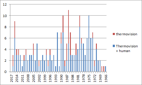
My attempts to appeal to the world mind give an ambiguous result: at the thermovision camera request, PubMed issues only 36 articles, with an amusing distribution by terms and specialties — cosmetology and dentistry prevail among medical fields by a large margin. Also, one group of neurologists from the Czech Republic published three articles on muscle pain, judging by the publication dates, we are talking about the same study, but splitting into three publications increases the proportion of pain studies in the overall picture. Not satisfied with this situation, I asked PubMed to search for “thermal” AND “camera” also in all databases.
In addition, I was curious how often these studies were applied to people.
It turned out that thermal imagers in particular were much more often used in the context of the study of people than methods of visualization of temperature regimes in general.
The issue of “thermal” AND “camera” turned out, of course, with a huge number of false-positive inclusions. I looked through the headlines and abstracts of the 500 most recent publications (total issuance of 830 points). As a result of this work, a lot of things were eliminated that in my subjective view are not applicable to clinical medicine - this list included articles on heat-conducting alloys, a lot of meteorological research, analysis of human recognition algorithms, nuances of electron microscopy protocols, and determination of alcohol content in beverages. I also referred to non-medical articles on the effects of laser acupuncture on the intestinal energy meridians.
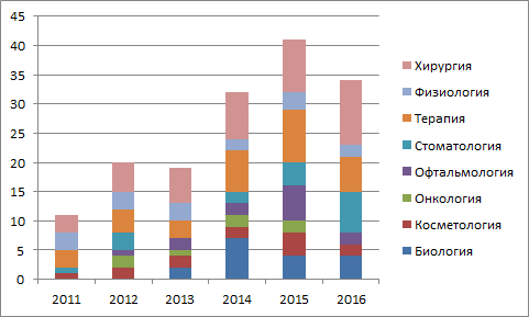
Among these five hundred publications were articles for the years 2010 - 2017. I dropped 2010 and 2017 as incomplete and made a schedule of the “popularity” of each of the topics in individual years.
I defined the subject of the article subjectively, but tried to adhere to more or less simple criteria. The topic of Biology includes everything related to the study of wild animals, veterinary medicine, and the meat and dairy industry. To the topic of physiology, I carried mainly experimental studies on one or another biochemical mechanisms in the experimental organism. As you can see from the graph, the main areas of interest are the same.
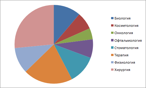
Here is the integral division by topic in the entire sample:
And here I would be happy for my colleagues - after all, various surgical specialties occupy an impressive part of the resulting “pie”, but I wasn’t sitting in one place again and I decided to check for what purposes temperature measurement is used in each article.
In this analysis, I removed from consideration of a group of articles on biology and physiology, as having no direct relation to clinical medicine, and also refused from dentistry, in which I understand almost nothing.
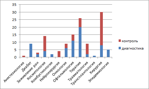
I marked the articles with the “diagnostics” tag, when temperature mapping indicators were used to identify the patient’s disease, or determine the stage of the disease, the location of the affected tissues in the body. The “treatment control” tag refers to those cases where the thermal sensor was part of the feedback mechanism: in real time, it evaluated the methods of treatment used - for example, it stopped working when the instrument overheated, or marked the treated areas of the skin.
The “treatment control” group mainly included articles on the use of laser in various fields - cosmetology, ophthalmology, neurosurgery, oncology, and many other places. On the methods of diagnosis was emphasized in the articles on diabetic defeat of the feet, various vascular diseases. Ophthalmologists have investigated the ability to diagnose dry eye syndrome. The popularity of epidemiological studies has come down to one question - the in-line measurement of passenger temperature at international airports. It seems to me that the topic is not as scientific as resonance.
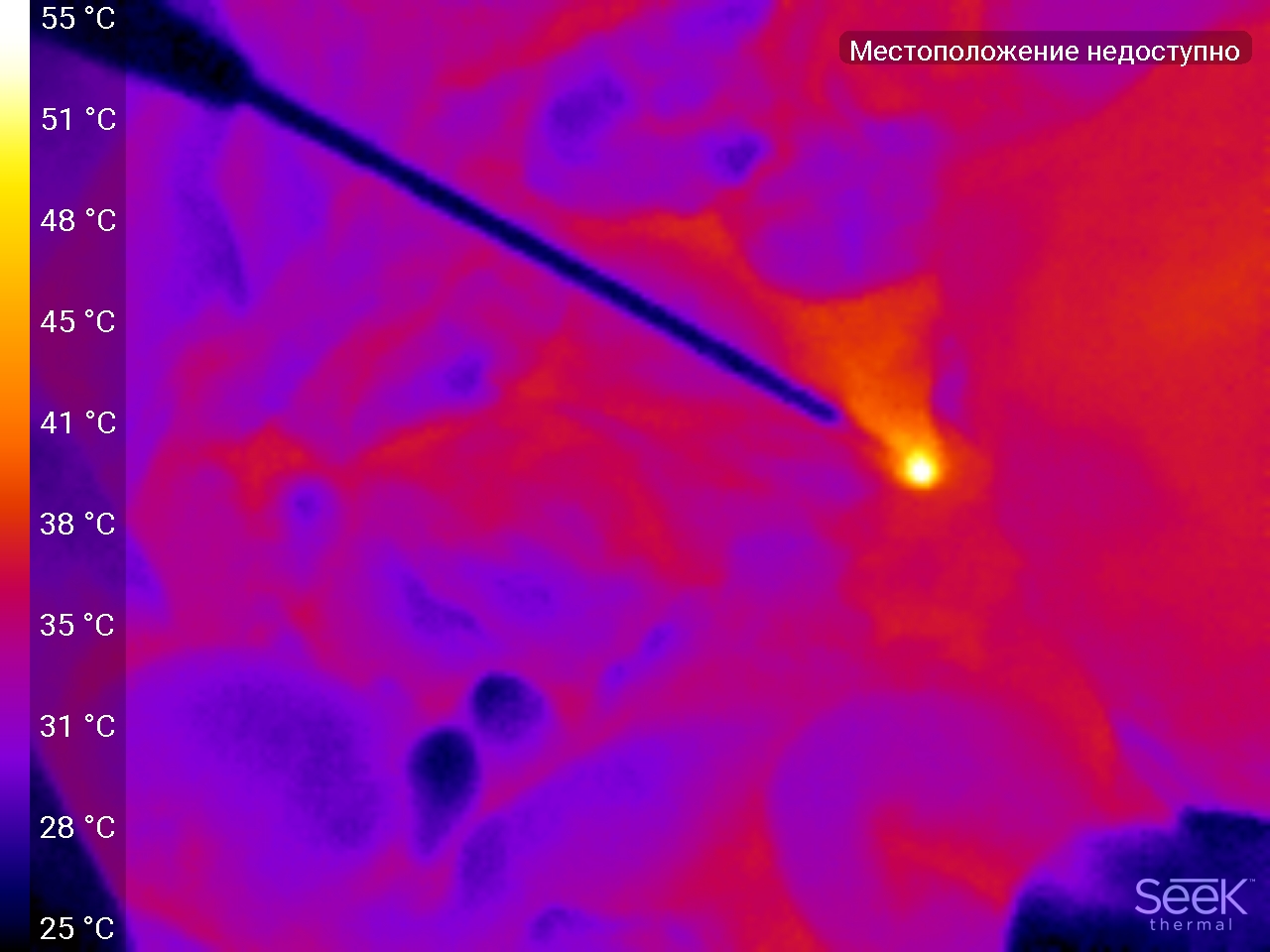
The share of "diagnostic" research in surgery is offensively small. In most cases, the authors tried to focus on the control of heating of surrounding tissues when working with various types of coagulating devices. How hot is it? Well, according to my measurements, something like this:
The black line is the tip of the coagulator, the white dot is the working surface at the very end of the tip + tissue that it touches. As you can see, with an average temperature of the surface of organs 31-35 * C, the coagulator heats up to 55.
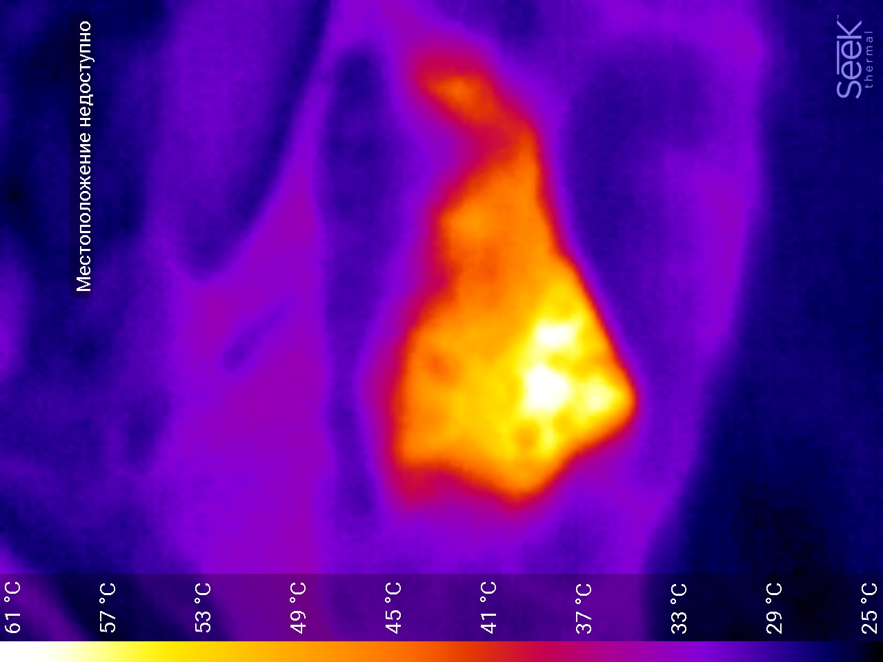
And this is what a portion of the greater omentum treated with coagulation looks like (diffuse bleeding was stopped).
Such temperature regimes are consistent with what is written in the articles. Good or bad - no one will say. At a distance from large vessels, working at such temperatures is quite acceptable. Fichey must be properly used so that there are no bugs.
EXAMPLES
I did not have a clear concept on what to shoot. Moreover, the results of my theoretical studies help only conditionally - in part of the articles they directly write that they used infrared radiation sensors, in part - that they used a professional thermal imager, and sometimes they don’t understand what the chip is all about - it’s only clear that they detected the temperature and made a conclusion from this.
In general, I was looking for what phenomena can visually demonstrate a snapshot of a thermal imager.
Hope to shoot everything and quickly disappeared in large numbers for two reasons: the laws of maintaining sterility are not always possible to get into the operating wound with a telephone, and technical - the technology of interventions is such that on the one hand the organs are in close contact and maintain each other's temperature and, on the other hand, we often use instruments that locally heat fabrics to high temperatures. If the first was clear in advance, then I could not imagine the scale of the second effect.
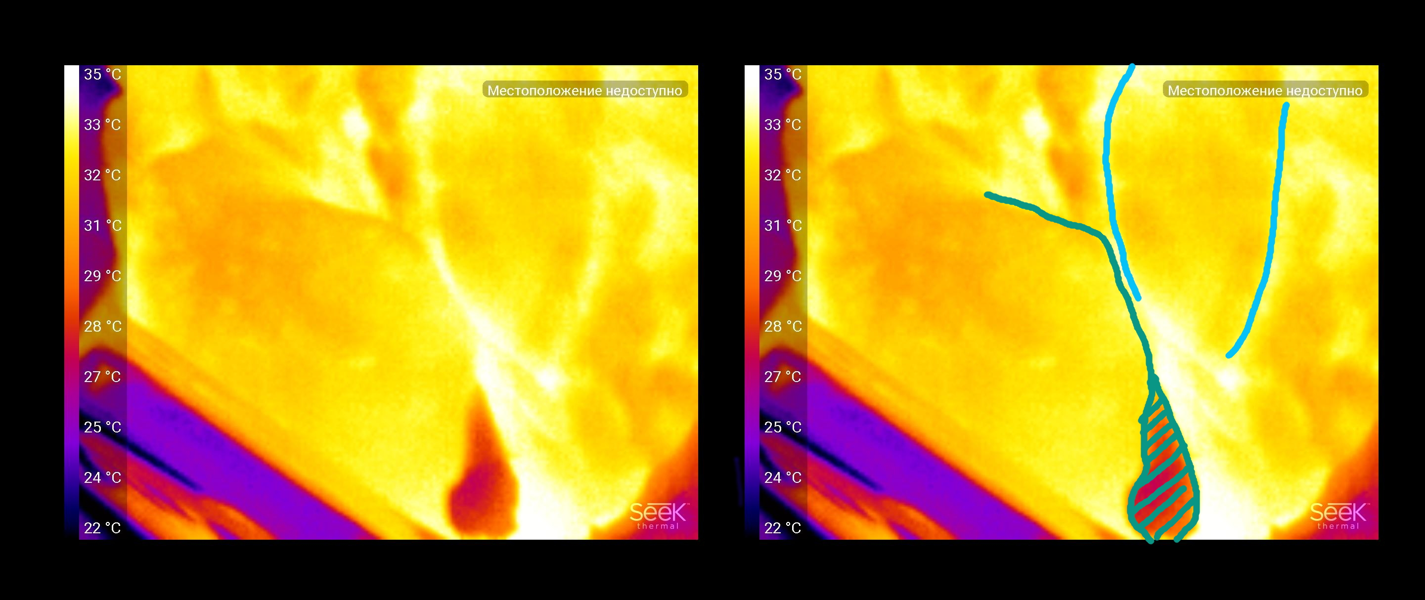
A bit norm
A healthy loop of the small intestine and mesentery with feeding vessels. 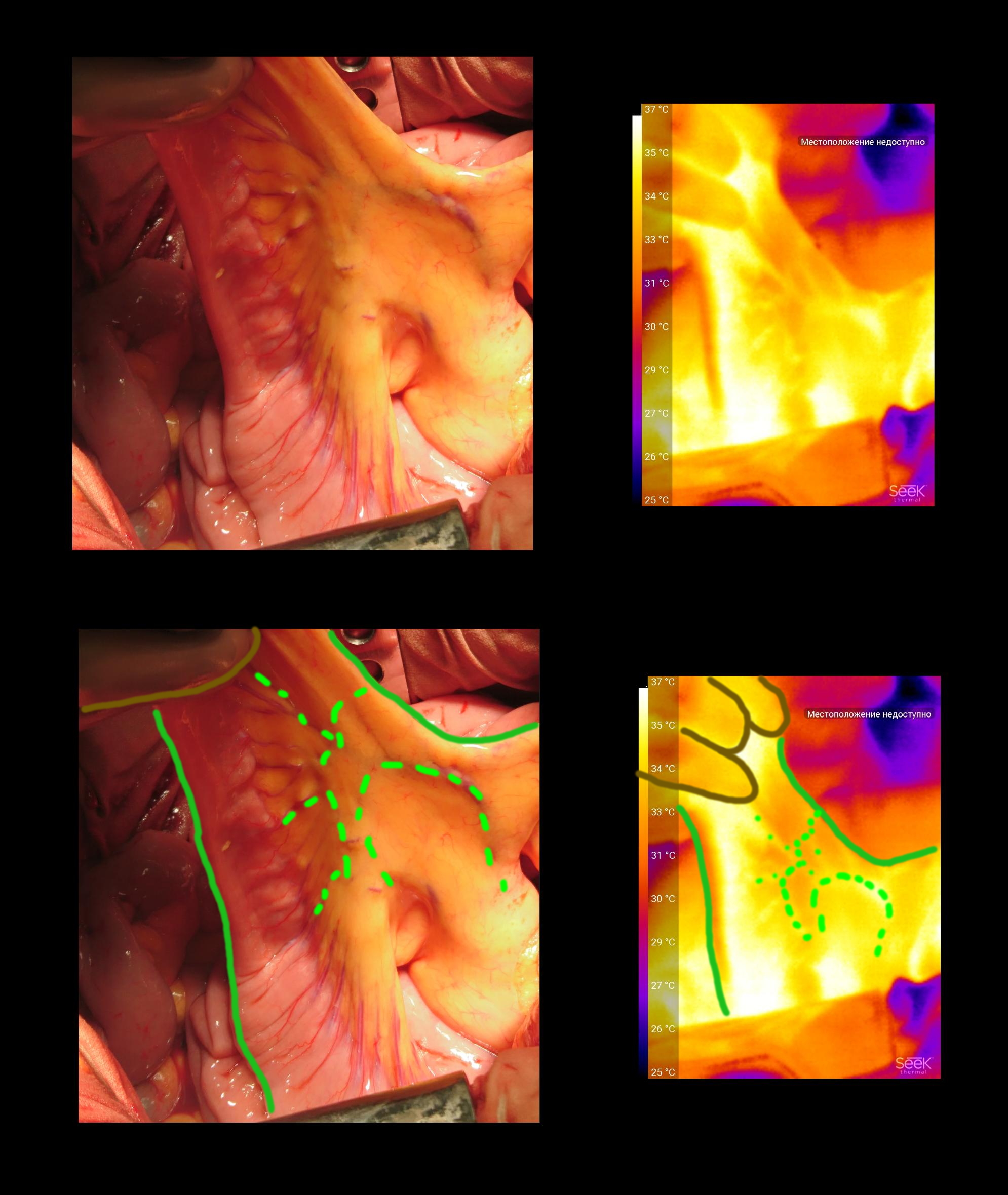
A healthy loop of the small intestine and mesentery with feeding vessels. The imager traces even thin vessels that are just not visible. In daily practice, these vessels are detected either when looking at the mesentery "for lumen" (badly helps with marked edema, lymph nodes, adhesions), or by touch by pulsation. Brown - surgeon's glove.
Colon after it was mobilized 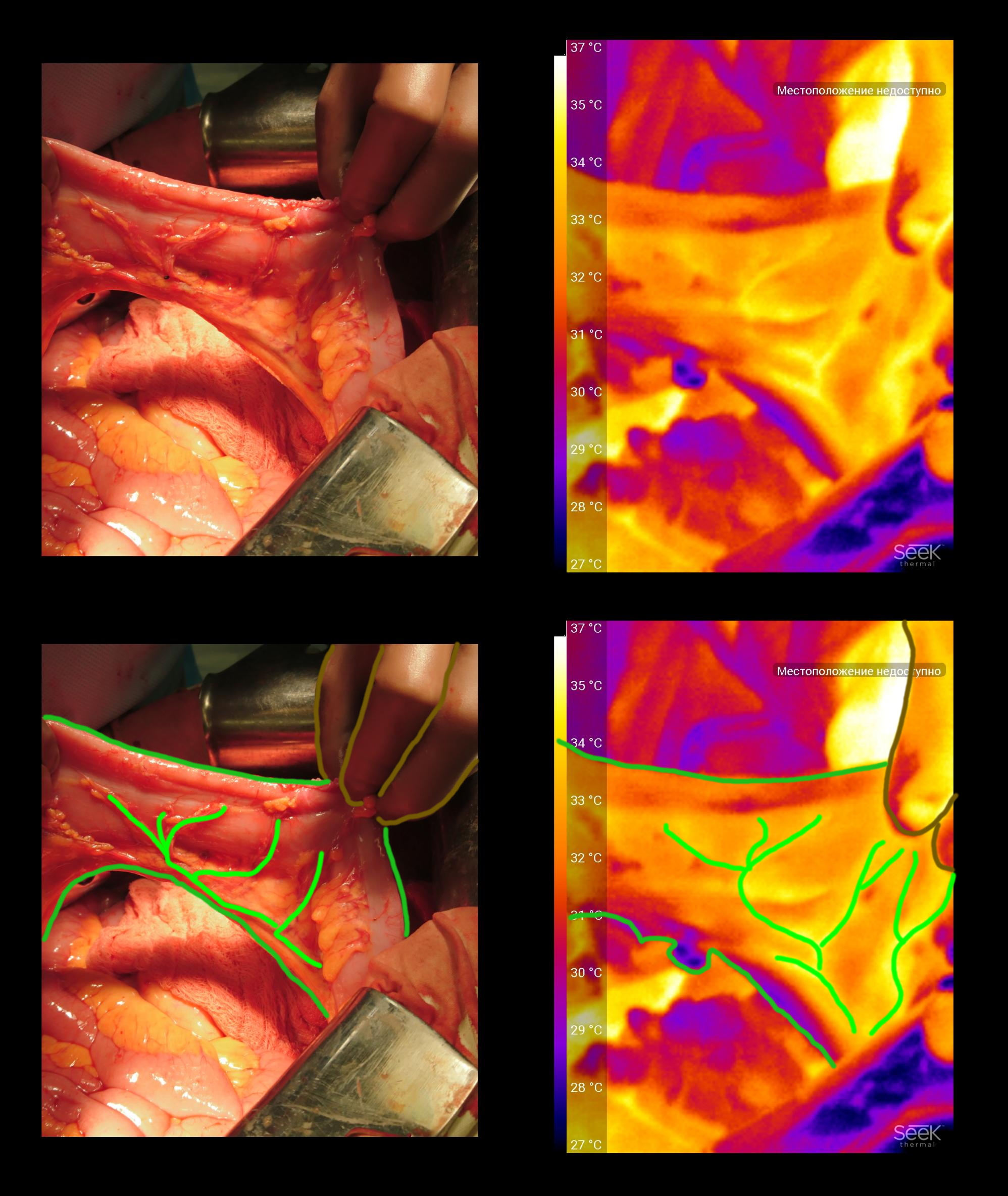
And this is the colon, after it was mobilized - freed from the ligaments that fix in the body. It can be seen that the feeding vessels feel very well and the intestinal wall is evenly stained on the thermogram - there is no edema or weakening of the blood flow.
Visualization of tissue viability during surgery.
The picture was taken during a stomach operation. 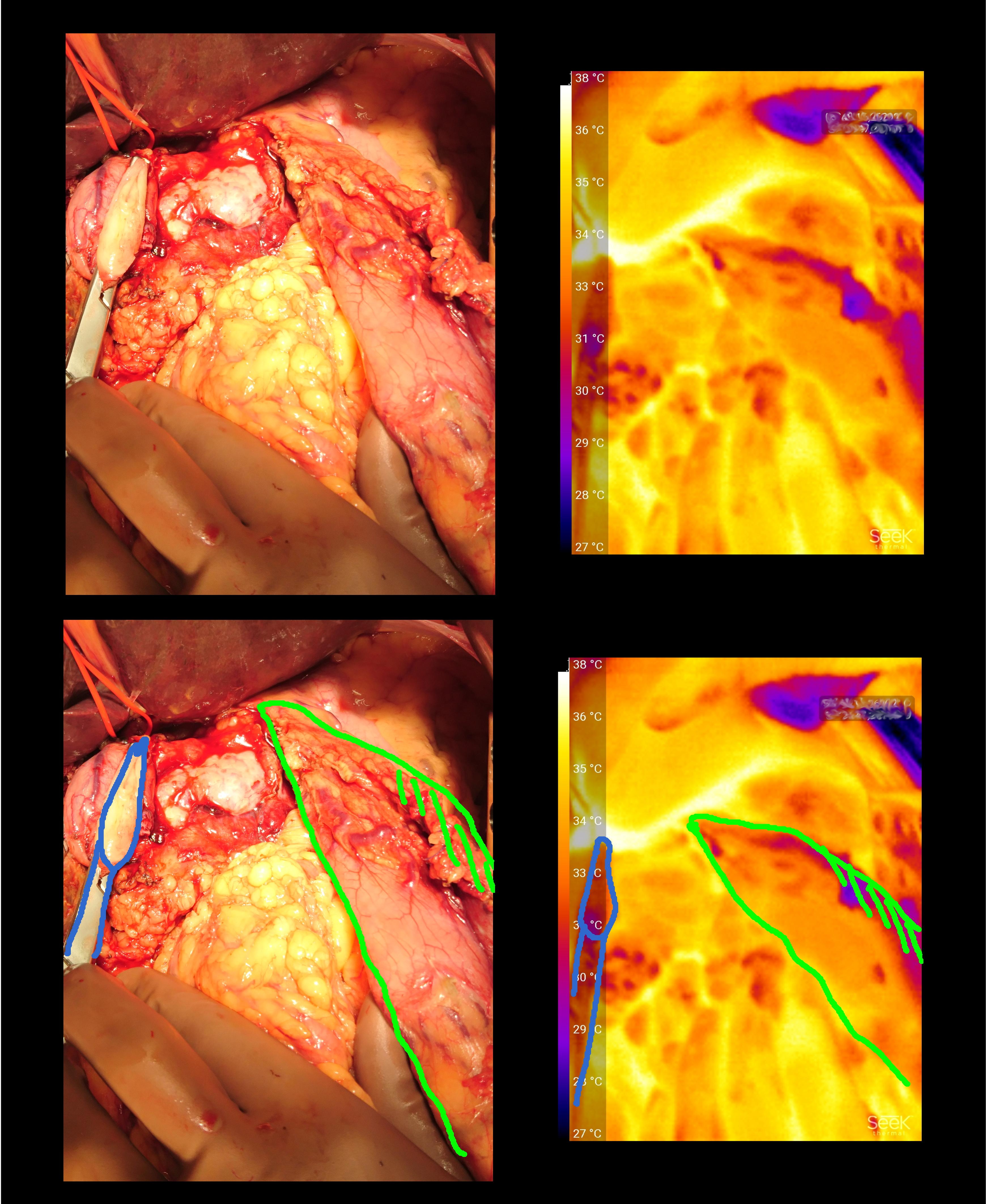
The picture was taken during a stomach operation. The remaining part of the stomach is visible on the right (indicated in green). It can be seen that the entire wall is uniform in temperature signal. The purple stripe (marked with green hatching) corresponds to the gland resection line - there the tissues are “sealed” with a coagulator and in the future they will form a scar.
Blue highlighted clip, superimposed on the duodenum. It can be seen that the tissues above the place of clamping are colder - they do not have blood flow, they will be removed during the reconstructive phase of the operation.
Another operation resection of the stomach. 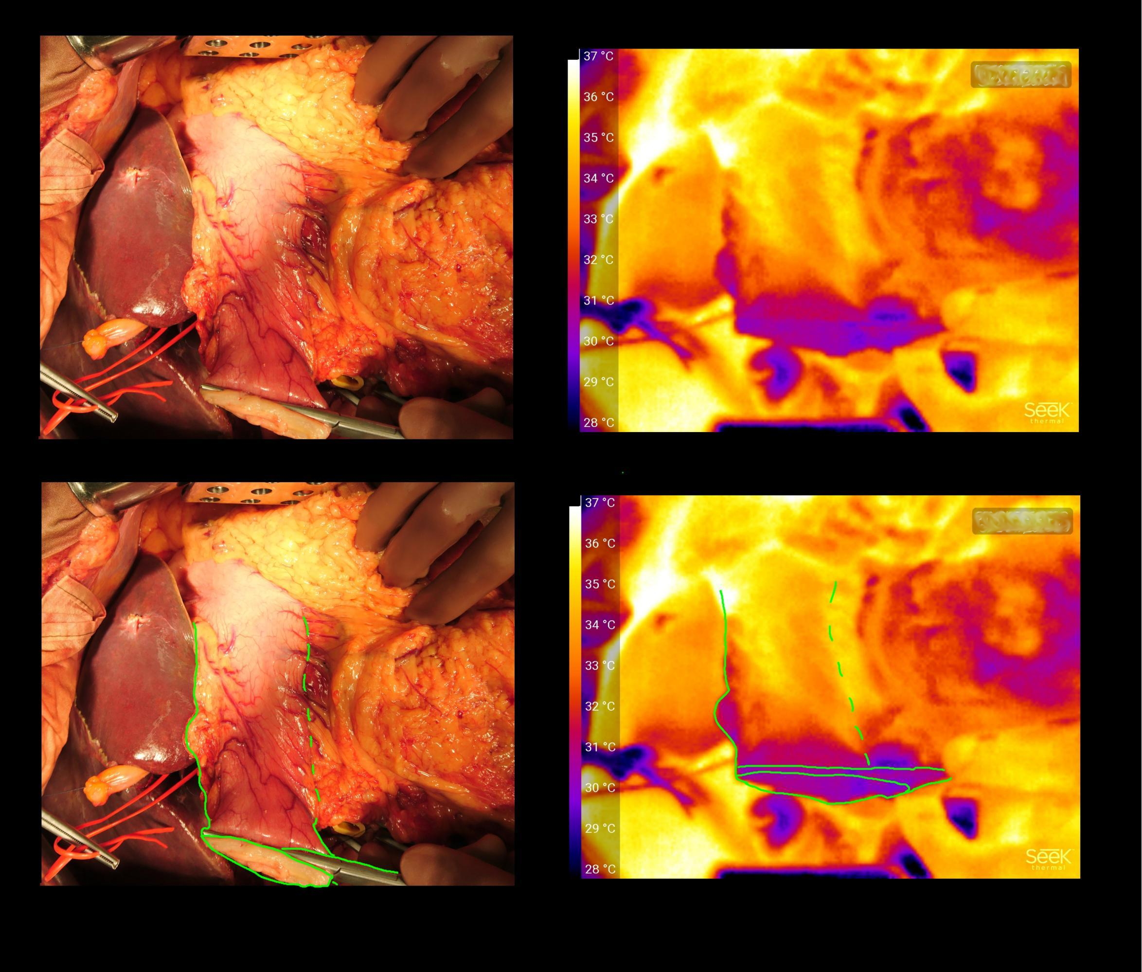
Another operation resection of the stomach. The color indicates the clamp on which the output section of the stomach, cut off from the duodenum. It can be noted, unlike the previous example, that the stomach sections close to the clamp are colder - the fact is that one of the arteries supplying this section is already bandaged.
Evaluation of postoperative suture healing.
Beauticians have long appreciated the benefits of temperature mapping for their specialty. As was shown above, more often the task of measuring temperature is the quality control of the procedure performed.
Nevertheless, judging by the results of my research, doctors use thermal imagers or infrared cameras to diagnose various conditions on the skin temperature - mainly neurologists shared this niche with the study of chronic muscular pain and sports medicine doctors who are interested in everything - and overworking muscles, and heat transfer in sports underwear, and, what intrigued me most, was scanning of the subcutaneous tissue with subsequent analysis of its thickness and biochemical properties.
But this is all the lyrics. In our department, it is possible to remove plenty of gradually healing postoperative wounds.
Here is the wound at the time of the end of the operation:
Wound 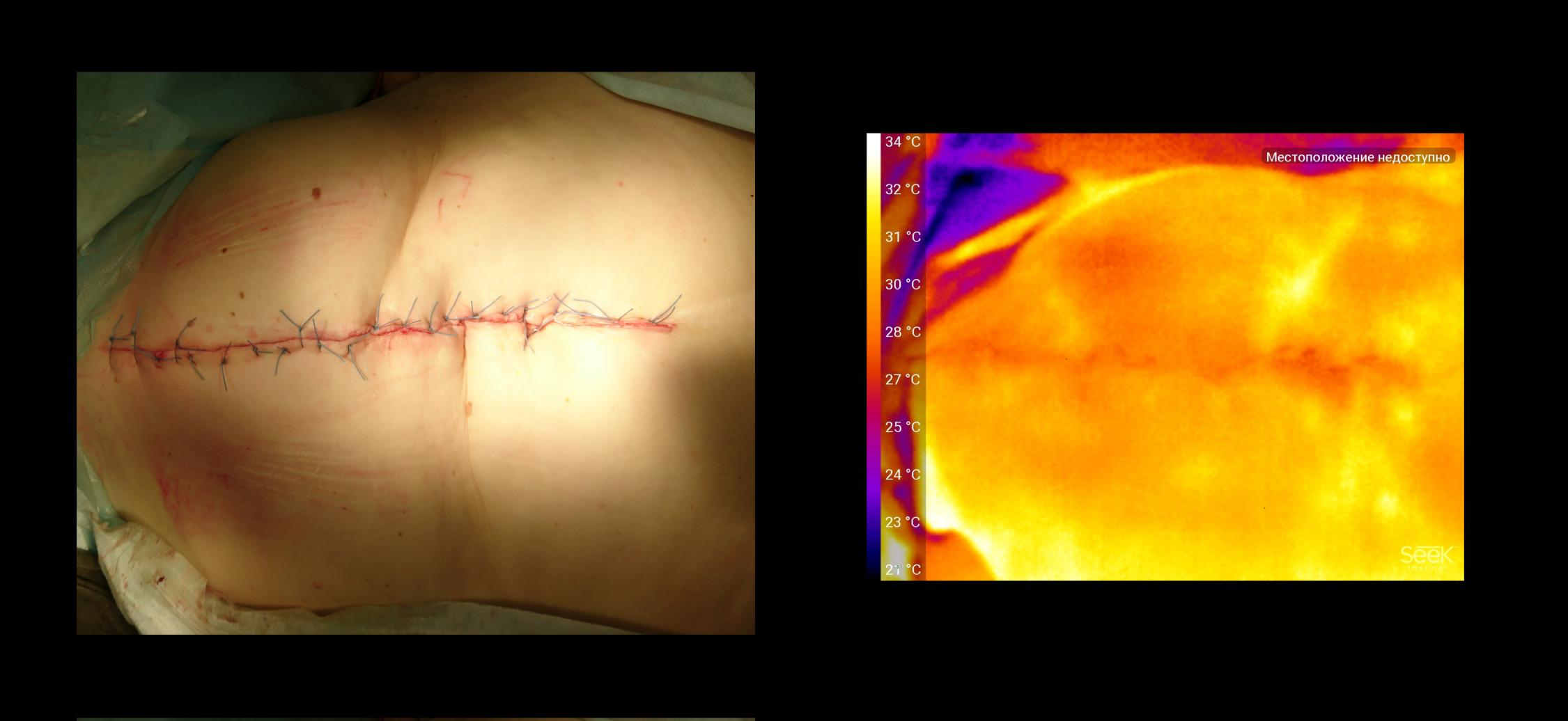
Some decrease in temperature is associated with dissection of the part of the vessels feeding the subcutaneous tissue and skin. In addition, the tissues exposed to the operation tend to swell (the nature of the edema is somewhat different from inflammation, while the vessels do not expand, but are compressed by the fluid from the outside), Additional ischemia (weakening blood flow) occurs because of the ligatures that form the sutures they further compress the tissue.
In addition to the factors described, the photograph illustrates the uniformity of ligatures, as well as the place of thinning of the subcutaneous tissue (oblique fold in the upper part of the photo). It exists for a long time due to the specific deformation of the spine of the patient.
10 days later 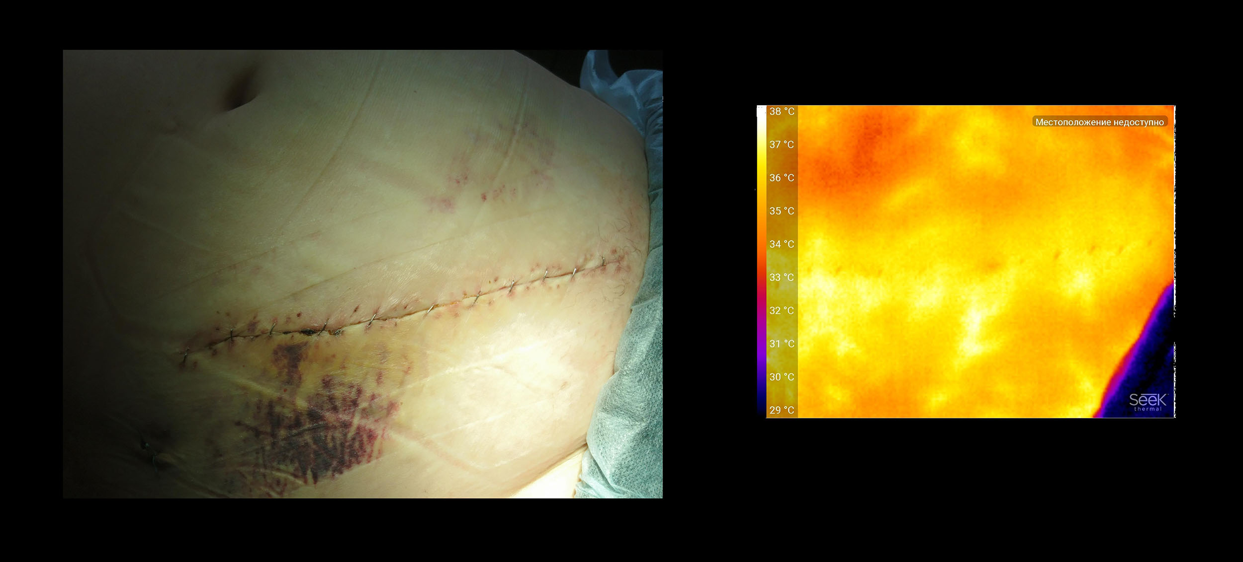
This image was taken 10 days after surgery. The skin was not sewn with ligatures but with special clips - they do not create ischemia. Thermal "patterns" on the skin at a distance from the suture, probably correspond to large vessels of the subcutaneous tissue. The patient received long-term therapy with steroids, a typical side effect is an increase in the body subcutaneous tissue in volume and an increased tendency of this fat to accumulate water in itself. Fat deposits are a good thermal insulator - they warm up slightly from deeper tissues, moreover, they produce very little heat, but the vessels against the background of such fiber are very contrasting.
I work in a planned surgical hospital without purulent separation. Inflammatory processes with the wounds of our patients are quite rare, and we are working to make it happen even less often. The patient from the next photo as one of the stages of the operation closed the colostomy - the exit of the colon to the anterior abdominal wall, which was formed a few years ago. Since the presence of a colostomy is a prerequisite for the inevitable contact of an operating wound with intestinal microflora, such an operation is called “conditionally infected” and according to statistics in such patients complications occur more often during wound healing.
Stoma 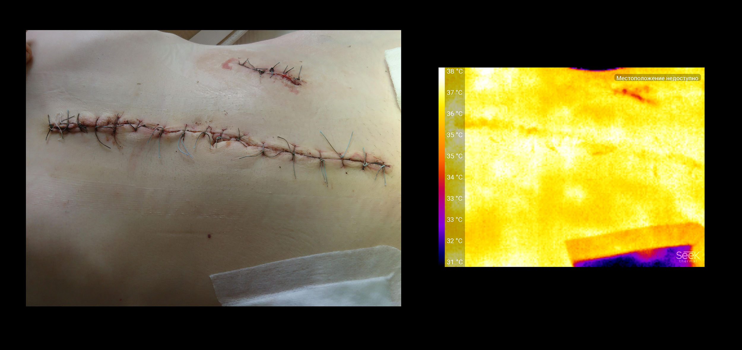
A large wound in the midline of the body is “clean,” it healed in normal time without any problems. It is sewn with ligatures - a certain decrease in temperature exactly along the seam line is noticeable (why this may be so - it has already been said above). The fact that the skin on the closest to the seam is 2-3 cm warmer than in other areas may be an artifact regarding the self-adhesive synthetic dressing - it creates a “greenhouse effect”, and the picture was taken immediately after its removal. But a small wound on the side, where the colostomy was, lags a few degrees behind. This is due to its “conditional infection” - a small amount of tissue that has come in contact with intestinal flora has lost its vitality, and in the healing process, these cells of the body “reject”. A pleasant discovery was the fact that foreign scientists statistically confirmed that in the presence of bacterial flora, the temperature of the seam drops.
CONCEPT OF APPLICATION: THE TECHNIQUE IS IMPORTANT THROUGHOUT ALL.
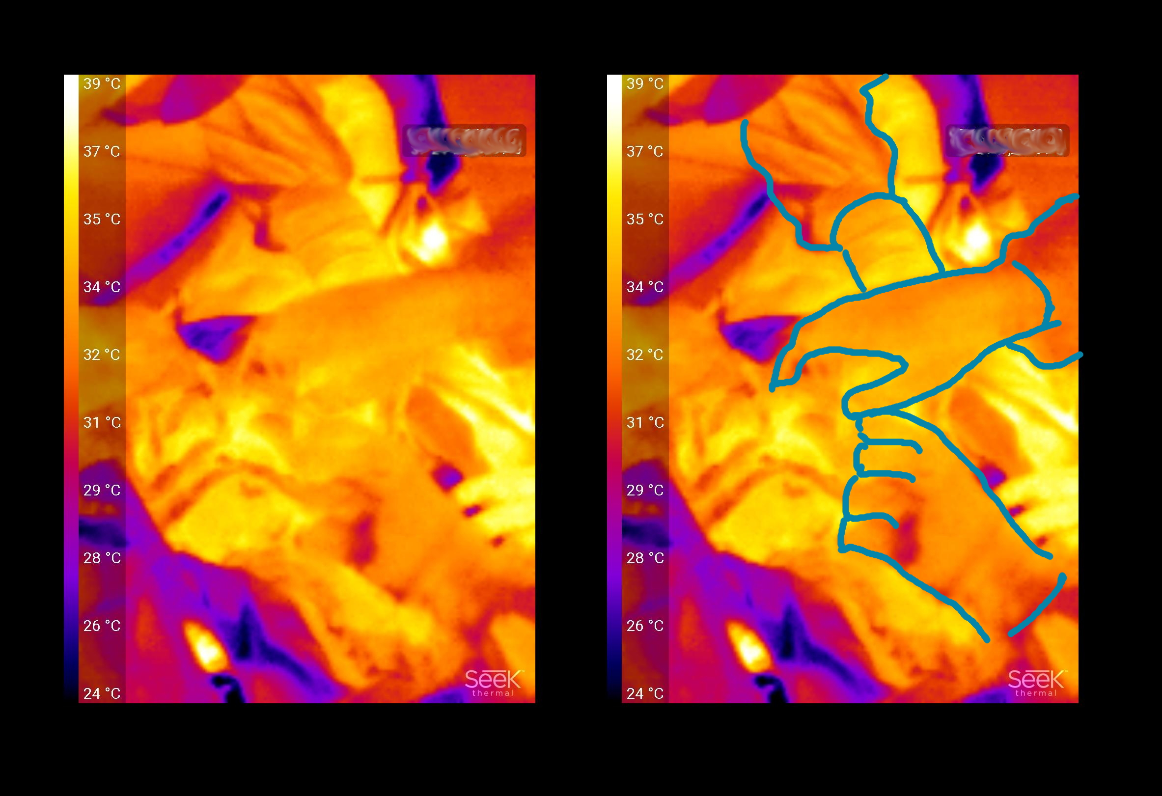
As I already said, my first attempts to photograph something were a failure. You cannot shoot with a thermal imager according to the same principles as with a camera. When photographing, I usually waited for the local work area to be clearly visible, and took a general picture - it was obvious who was doing what and in what area. With a thermal imager, this number does not pass - the surgeons' hands do not contrast with the patient’s organs.
A typical situation when I asked everyone to remove hands, napkins, tools from the wound and quickly filmed what happened:
In fact, this frame has a rather large surface of the liver (left), the output section of the stomach (highlighted on the right), the small intestine is the right edge of the frame. Everything except the gallbladder (hatched) merges.
The next step was to “hold on the weight so that it was visible”. Organizational problems are already connected with this - not only I (the assistant) are distracted to take off gloves, take pictures, then wash my hands again, put on a new set of dressing gowns and gloves, but the operating surgeon has to postpone work and help me. And still, success is dubious.
Failed 
But it turned out to be a problem to be solved. It was necessary to somehow neutralize the effect of heating from neighboring organs - then the picture will correspond to the tree of blood vessels. For example, wrap the organs marked for a photo in a napkin moistened with a solution of room temperature. The number of visual frames has increased significantly.
The idea was not new, the dentists had already described the method for determining teeth cutting out in children, when the oral cavity was first blown with cold air, and then the temperature of the gums were recorded.
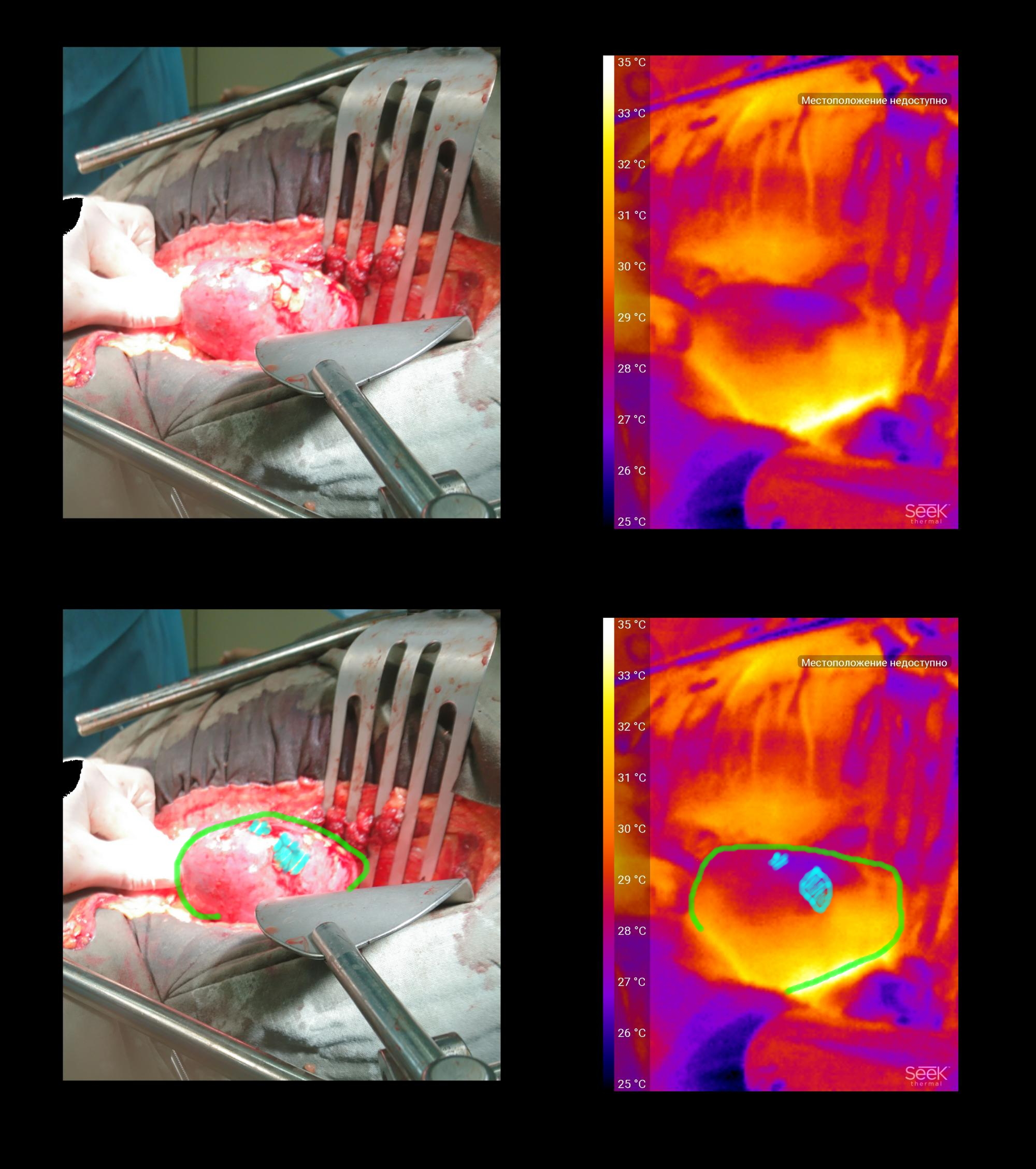
In my case, the methodology has led to a very clear visualization of a colon tumor.
Tumor 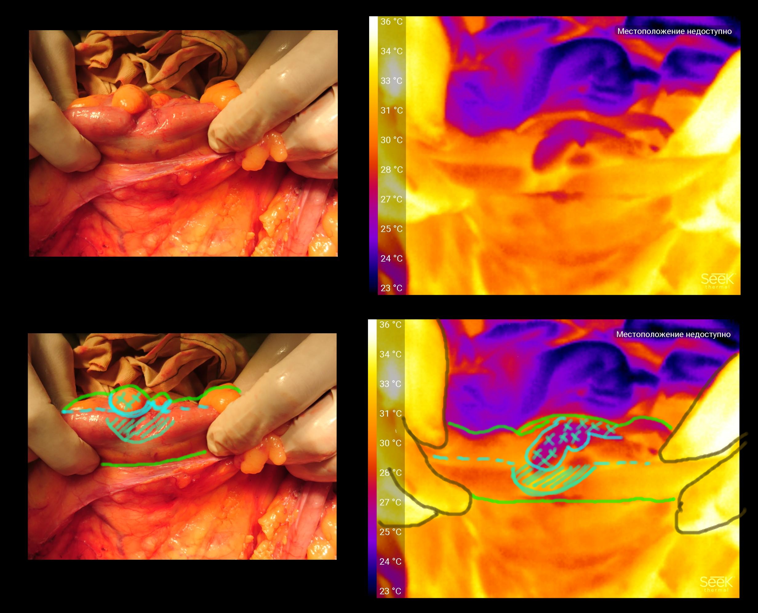
The tumor grows inside the intestinal lumen, but it deforms the wall outside (shaded area). In general, the tumor tissue is characterized by a higher level of metabolism, which is why it is lighter on the wall above the tumor. But because of the swelling around the tumor, the blood supply to the fat suspension of the intestine (filled with blue crosses) suffers, it is much colder than its neighbors.
MEGA BONUS
In the issue I analyzed with PubMed there was one article from the transplant section. In it, the temperature of the surface of the abdomen tried to determine the level of metabolism after bowel transplantation. I suspect that many technical details awaited the researchers on this path, but I’m talking about something else. Transplantation of the intestines is not limited, and the assessment of the adequacy of the blood supply there is very important.
Most often, the blood supply of the graft is estimated by its color - during the preservation period the organ is washed with a colorless solution, therefore the tissues become yellow-brown-gray. And after the start of blood flow stained in the range from pink to red and lilac.
Liver 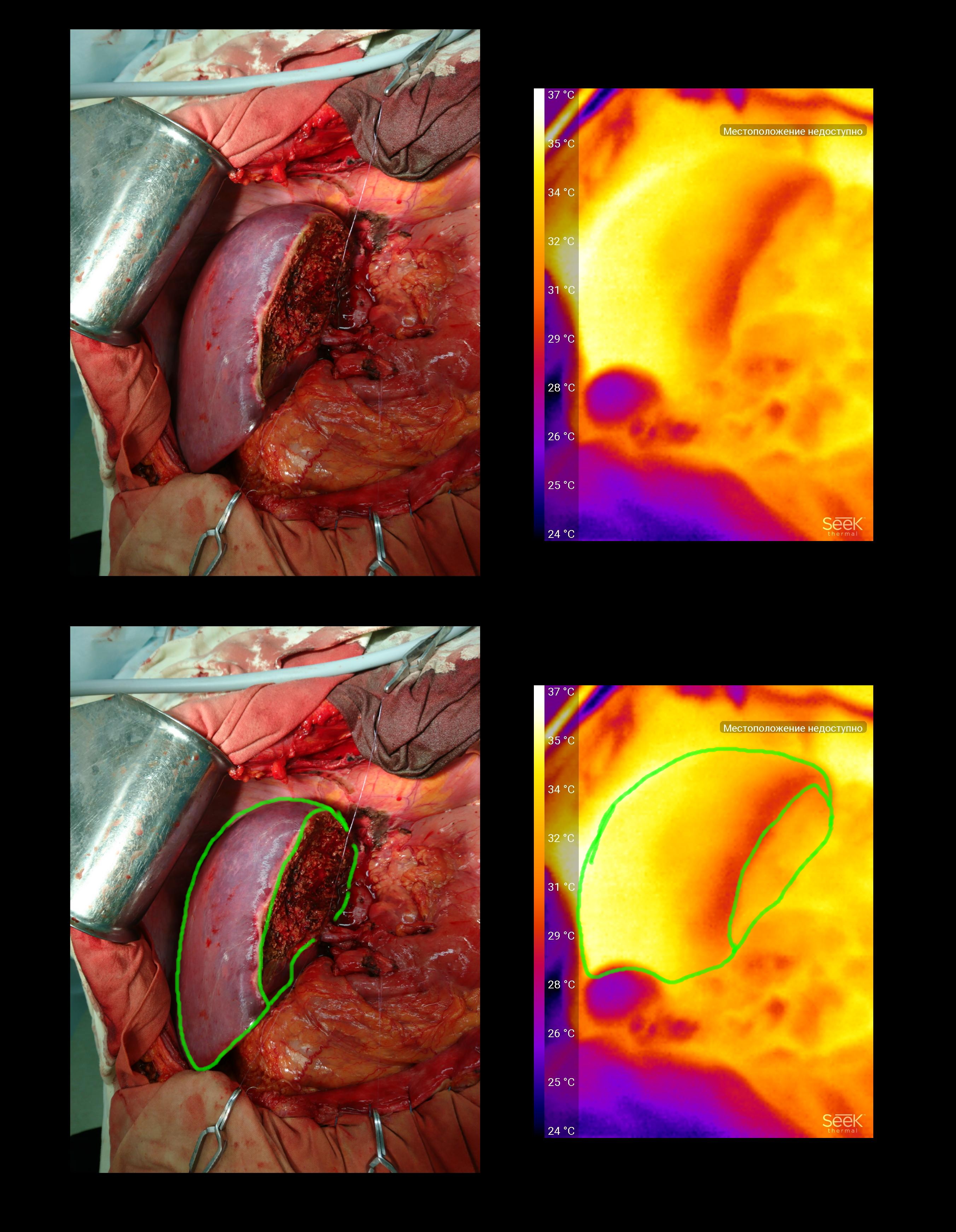
The graft is half the liver. After starting the blood flow acquired the correct uniform color.
The thermal imager shows that the tissues located under the organ capsule along the resection line are worst of all blood supply, which is quite logical, since during the work there was a thermal damage to the blood capillaries and the tissue itself. The fact that the right segments look much warmer is an artifact, they warmed up from neighboring organs. In this case, given the uniformity of color transition, we can say that the perfusion is adequate.
Liver 2 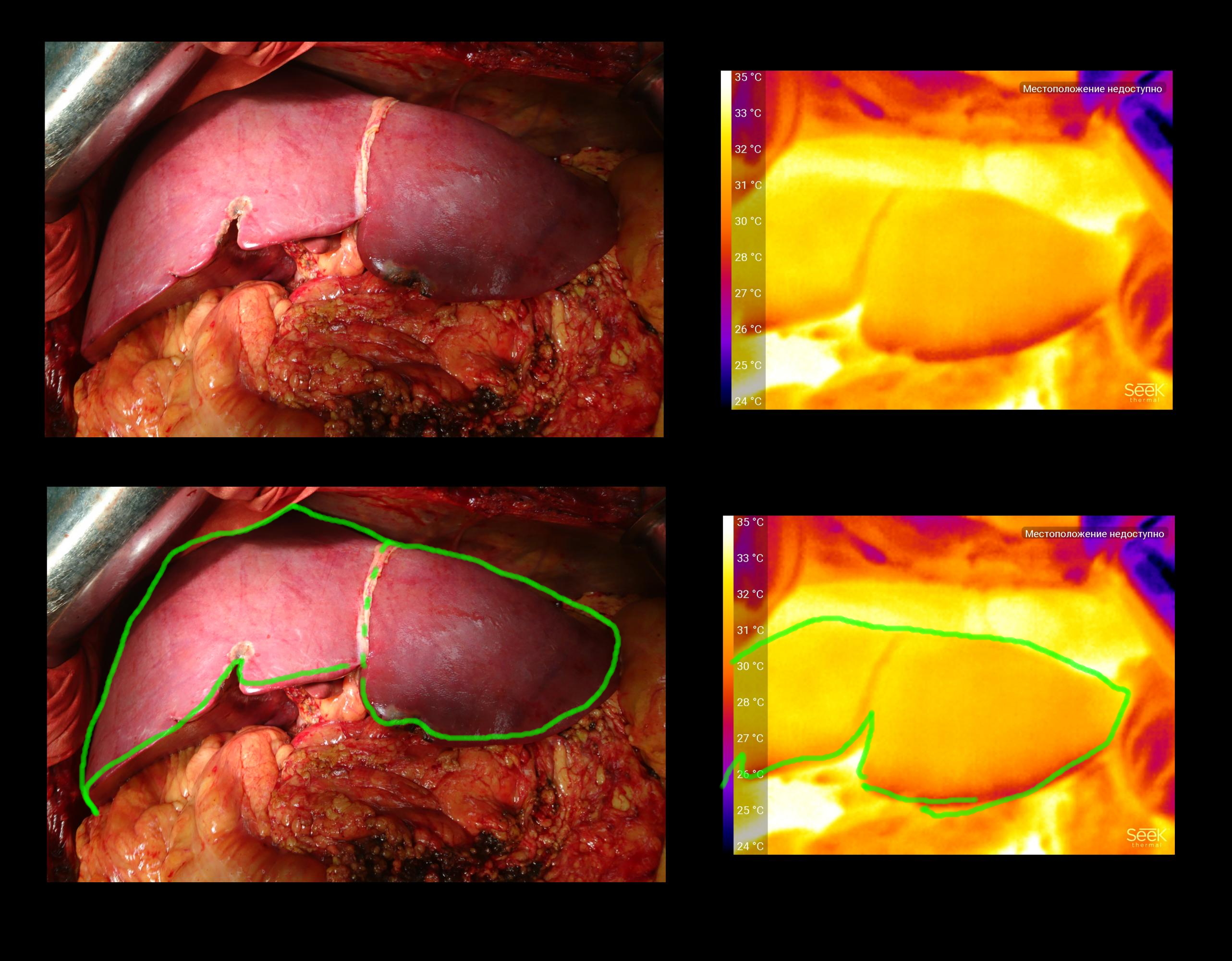
The graft is a whole liver. Was preserved for several hours. Coloring is also uniform.
Bud 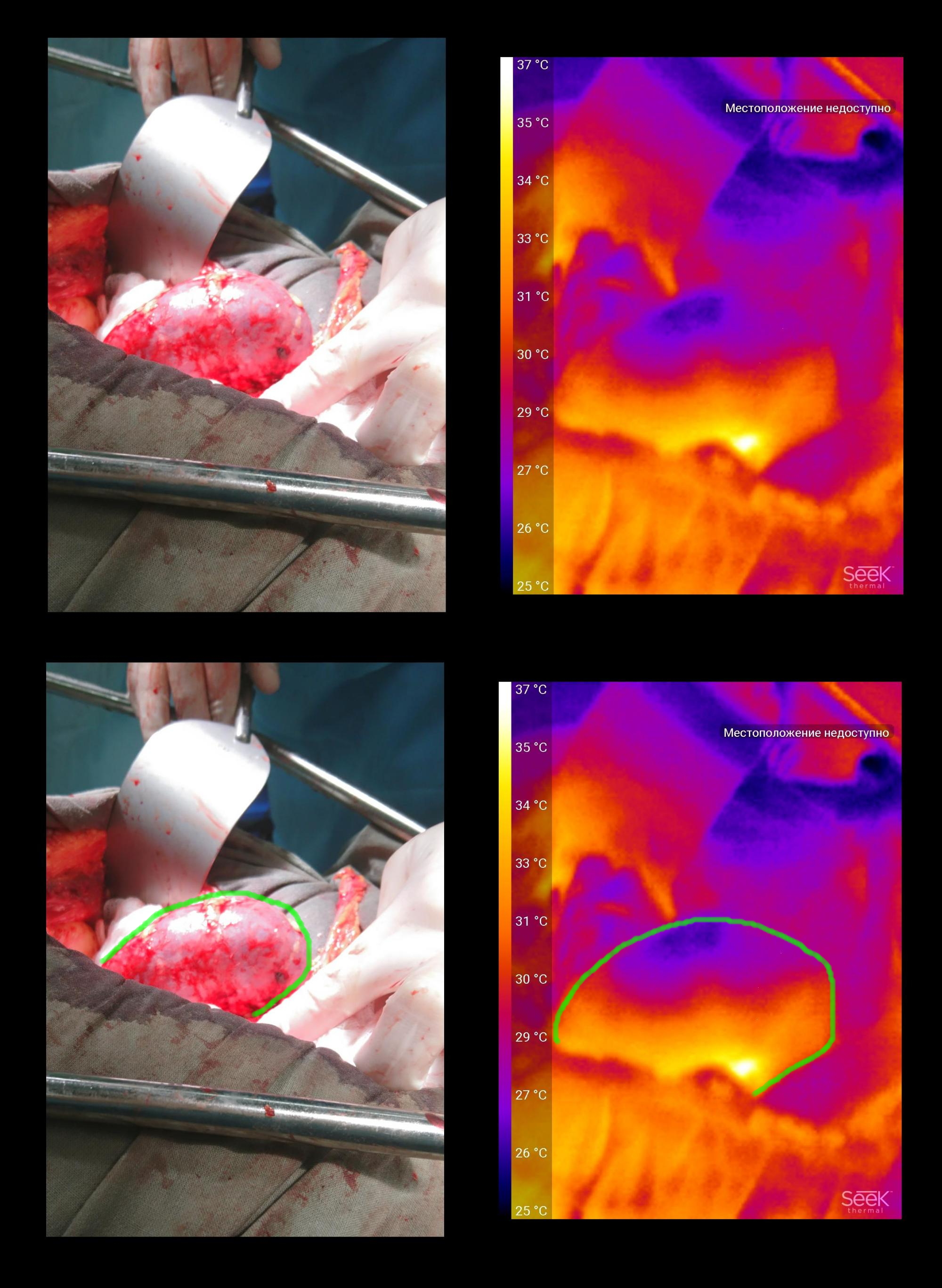
The kidney transplant, the picture was taken immediately after the start of blood flow. It can be seen that the temperature in the “organ gate” area is restored first, where the vessels flow. In the picture it is at the bottom of the kidney. Blue marked several fragments of kidney fat - it does not supply blood from the renal vessels, so it remains colder.
Sometimes, to cause reflex relaxation of the vascular walls, the graft is easily massaged. The video shows how after that the temperature of the entire surface rose (the tissues warmed up a little by hand), but then the kidney itself retained this temperature due to improved blood flow, and a piece of fat cooled to its original state.
FINDINGS
Unfortunately, the revolution “came, photographed, made a diagnosis” did not work.Of course, the majority of routinely used methods (assessment of tissue color, tone, peristalsis) are subjective and appeal to the doctor’s experience, but at the same time, the principle “if you doubt the viability of tissues, consider that it is not viable”. And hardly anyone agrees to preserve fabrics that look dubious outwardly, even with normal indicators of technology.
I would like to share my impressions into several areas:
Ease of use in the operating room is low: first, the device cannot be placed in a sterile casing, therefore either you need to ask someone (who doesn’t understand very quickly what it is), or wash your hands several times, which spends a lot of time, gloves, gowns. Secondly, it is difficult to direct the camera exactly where it is necessary - the beam is narrow and does not come from the place where the camera is usually located. The latter is partly solved by turning the phone over, but not that this is the perfect solution. The most organic solution I personally see is an aquabox-type casing that covers everything except the lens, a Micro-usb extension cable, so that the phone can lie to the side.
The second block of impressions concerns the medical side of the issue. An analysis of the literature showed that the direction of temperature mapping in medicine is promising, but it requires elaboration of the methodology (such as the idea of pre-cooling the subject), and needs an “ecological niche” - a field of application where mapping would open up new possibilities. There are examples of successful solutions - diagnosis of trophic disorders in patients with diabetes. I personally wonder if it is possible to evaluate the functioning of the graft through these methods. If due to such a non-invasive and conditionally cheap procedure, it would be possible to more accurately select therapy in the postoperative period, it would be very cool!
Many thanks to my husbandwho agreed on a thermal imager and edited the article, and colleagues for their responsiveness, patience and assistance in filming.
The SeekThermal thermal imager was provided by the company Dadzhet, the official distributor of SeekThermal in Russia. Dadjet suggested that I publish the promotional code Thermal Imager for the readers of this article, which gives a 10% bonus on its purchase.
All Articles