Stanford neural network diagnoses pneumonia on X-ray better than doctors
Researchers at Sanford University have developed a self-learning algorithm that can diagnose medical imaging (chest X-ray). This software platform specializes only in pneumonia, but it performs its task better than professional radiologists .
Pneumonia is different, and the neural network is distinguished by its 14 varieties on x-ray. The results of their work, scientists have published in the public domain on arXiv . “Analysis of medical images is a difficult task, and we know about it,” says one of the developers of the software platform. “We decided to develop self-learning algorithms by conducting a“ training session ”based on hundreds of thousands of medical images.”
The work uses the data set provided by the NIH Clinical Center. This is a huge database, which includes more than 112 thousand frontal images of the human chest, obtained by fluoroscopy. In these pictures, you can distinguish 14 different pathologies. This information made it possible to “pull up” a software platform for diagnosing these pathologies.
After the training was completed, the scientists decided to check how well the system makes the diagnosis. After the car was diagnosed, the researchers asked the doctors to do the same. Doctors analyzed 420 images and made their own diagnoses for each of them. As it turned out, the computer system was able to diagnose pneumonia more accurately than humans.
Pneumonia is a dangerous and common disease. In the United States alone, with pneumonia, about 1 million people get to hospitals every year. Some pathologies are extremely difficult to detect on X-rays - too implicit signs in some varieties of this disease. At the same time, machine learning can help doctors cope with this problem.
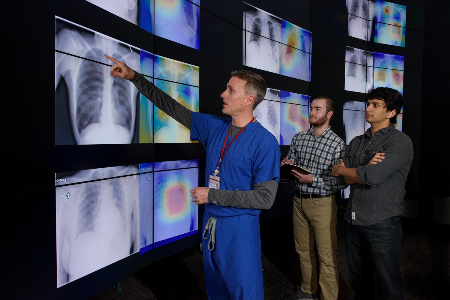
The main danger for the patient here is that if the doctor incorrectly diagnoses, it can lead to the appointment of the wrong treatment. As a result, the patient will be treated, but his / her condition will begin to deteriorate. The situation is aggravated by the fact that doctors have to analyze hundreds of images of this kind per day, as a result of which attention is dulled. A machine, provided its high-quality training, could work around the clock, almost without making mistakes. Moreover, the computer system can highlight in the photo the smallest details that are important for establishing the correct diagnosis, but imperceptible to humans.
The developers "taught" their system to create when analyzing images of something like a thermal map of the human body. Only instead of showing the temperature, parts of the lungs are marked with different colors, where the car “saw” signs of pneumonia. After processing, the pictures are examined by a person, paying attention, first of all, to those areas that the car has marked as the hottest.
Researchers believe that such systems will soon become generally accepted and common. “We are going to continue to create and improve medical algorithms, auxiliary to detect deviations from the norm. We also hope that we will soon be able to share depersonalized medical data sets that can be used by other specialists working on similar or other problems, ”said Jeremy Irwin, a representative of the research group.
X-rays in medicine - the most important source of data on the patient's health. Many doctors are simply inundated with similar images. After several hours of working with them, the ability of the doctor to concentrate decreases, attention decreases, so the probability of error is high. Automating the process could help solve the problem, which would reduce the number of medical errors.
The work carried out by scientists is not unique. Now many startups and large corporations like IBM and Google are engaged in similar developments. In addition to pneumonia, computer systems already know how to detect signs of tumors, cardiovascular problems and other deviations from the norm on X-rays and other medical images.
All Articles