Astigmatism - vision as in the kingdom of "crooked mirrors"
Friends, after a break, we resume publications on vision and technology to restore it. The pause was connected with my workload of operations and participation in conferences: in the near future I will publish a review of the most interesting technical innovations in ophthalmology that were presented at them, and today we will talk about astigmatism.

Modern statistics depressing. More than half of the world's population suffers from visual impairment, the most common problems are myopia and hyperopia. But ophthalmologists often diagnose another disease, the name of which is unfamiliar to many. Astigmatism is a defect in the optical system of the eye when the sharpness of the image is asymmetrical in vertical and horizontal. And the parallel rays of light passing through the eye are not focused into a point, but into the “eight”. For a person, this means that the visibility of the image becomes unsharp, and this often applies to both distant and close objects. As a result, instead of a normal image, a person sees a distorted image, in which some lines are clear, others are blurred. An idea of this can be obtained if you look at your distorted reflection in an oval teaspoon. A similar distorted image is formed during astigmatism on the retina.
Ophthalmologists say that almost two thirds of the world's inhabitants face this problem. But since the degree of astigmatism may be small, many people practically do not feel any discomfort. It is difficult for doctors to identify a clear list of common symptoms indicating that a patient has astigmatism. In each case, they will vary. In the very early stages, it is often confused with tired eyes.
However, quite a lot of people need special treatment or correction of this disorder with the help of glasses, contact lenses or even surgery.
Astigmatism is a defect in the optical system of the eye. In this case, the refraction (or reflection) of the rays in different sections of the transmitted light beam is not the same.
As a result, the image of objects becomes blurred, each point of the object is displayed with a blurred ellipse. And the whole picture, like in a “crooked mirror”, turns into a so-called figure “Sturm's conoid”. In practice, the main complaint is blurred vision at a distance, often vague near blurring, vagueness and division of objects.
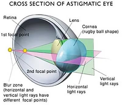
The course of the rays in the eye with astigmatism looks like.
This is the first question that patients ask when they hear this diagnosis for the first time.
Astigmatism may be congenital or acquired, may be stable and progress.
Quite often, astigmatism is a feature of the structure of the cornea at birth, it can even be inherited from parents. If its value affects visual acuity (as a rule, we are talking about astigmatism greater than 1 diopter), then it necessarily requires correction from an early age. Otherwise, the eye sees poorly and develops amblyopia, the “lazy” eye, which is no longer capable of high vision in adulthood. A child with astigmatism, by the way, will never complain to you about poor eyesight - he doesn’t know how to see this well. Therefore, early examinations by an oculist and wearing glasses, "as prescribed by a doctor," is so important.
It often happens that a grown-up person is surprised to find out that he has, as it turns out, astigmatism, this is in the case of those not examined in childhood.
Astigmatism may appear already in adulthood as a result of corneal diseases, such as keratoconus, inflammation, trauma and operations.
In any case, it does not appear from working at the computer.
First of all, the cause of astigmatism can be two optical systems of the eye - the cornea and lens. When glasses or lenses are picked, they take into account the resulting optical power of these two systems. Please note, patients usually know about frequent corneal astigmatism, but forget about the lens - it can make a difference, for example, in cataracts, when irregularities appear in the lens. "The cornea is flat, but there is astigmatism" - this is it. Often the patient and the optometrist do not bother at all with what is the cause. When selecting points, it is important to correct the rays of light onto the retina using correction. And with surgical correction - this is important.
For a simplified understanding of the essence of the problem, I often explain to patients that the optics of the eye with astigmatism is not like a sphere (a football), but an oval (a rugby ball).

First, the value of astigmatism matters - conditionally up to 1 diopter, astigmatism is considered physiological, weak. That is, as a rule, it does not require correction. But in case he even lowers his vision, alas, he needs to be corrected. The average degree - up to 2 diopters, high - 2-3 diopters and more than 3 diopters - this is a very high astigmatism. There are other classifications, but this one best reflects the effect on defocus.
Secondly, there are two types of corneal astigmatism: regular and irregular.
With regular astigmatism, there are two main refractive perpendicular cross-sectional planes, within which the optical power does not change - weak and strong. In such optics there is at least some regularity, even if the axes are skew or there is a big difference in their optical power.
Regular astigmatism - in this case, two parts can be distinguished, where the cornea has a different degree of refraction. This type of astigmatism can be corrected with glasses with cylindrical glasses or soft contact lenses.
Irregular astigmatism - in this case, two parts cannot be distinguished, since there are many optical axes. This type usually develops as a result of corneal damage, for example, during an accident (scar tissue forms), or due to uneven corneal protuberances (keratoconus). Irregular astigmatism is usually poorly corrected, sometimes hard contact lenses or laser effects on the cornea can be useful.
Third, the type of astigmatism depends on the location of the strong axis. If the strong axis is vertical or no more than 30 degrees deviates from the vertical - direct astigmatism. If the strong axis is horizontal ± 30 degrees - astigmatism is reversed. If in between - astigmatism with oblique axes. Direct astigmatism slightly increases the depth of focus, the reverse - more impairs visibility, but these both options are well corrected. But the correction of astigmatism with oblique axes with glasses and lenses is worse performed.

In astigmatic eyes, there are two perpendicular to the section plane with the greatest and the smallest refractive power.

Fourthly, depending on how the focus relates to the retina, astigmatism is divided into myopic (myopic), long-sighted (hypermetropic) and mixed - when one part of the image falls in front of the retina and the part behind the retina. For example, minus two and minus four - then this is complex myopic astigmatism, plus two and plus four - complex long-sighted astigmatism, plus two and minus four - mixed.
Nearsighted astigmatism is indicated by the sign “-”, long-sighted - by the sign “+”, when mixed in the eye there are “+” and “-”.
Symptoms of astigmatism are a decrease in vision, sometimes the vision of objects is bent, their split, rapid eyestrain at work, headache. Often you may not even suspect that there is such a problem, complaints are so typical - poor vision for distance or near, or at all distances.
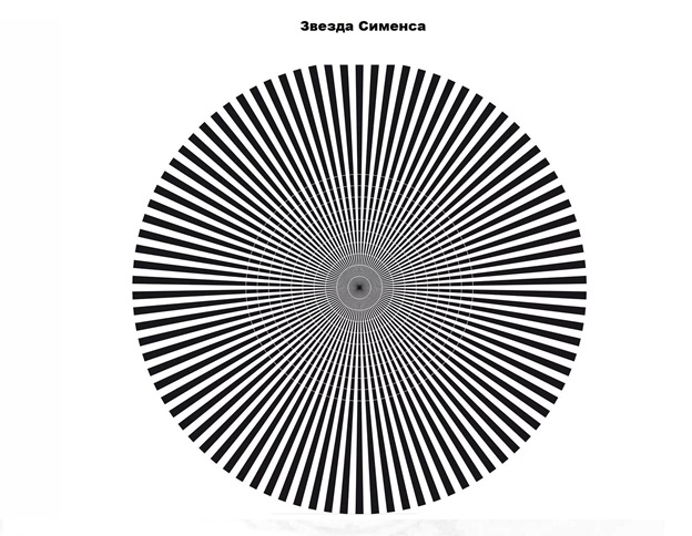
Since astigmatism is a defect in optics, then, firstly, the optics of the eye are carefully examined. And the correct conclusion will give only an ophthalmologist. To do this, we have a series of tests: a study on an auto-refractkeratometer, an aberrometer, a phoropterer with the selection of test lenses to determine visual acuity with maximum correction. A very interesting radiant figure gives an idea of how much astigmatism is present - if you look at the center of the figure from below, some of the lines will be clearer, and some will "blur" with those who have astigmatism.
In the phoropter tests, an ophthalmologist in the arsenal also has a number of techniques to clarify the presence of astigmatism, its axis and size. And this process is always quite time consuming - this can be confirmed by those patients who have done with us, for example, SMILE. Verification of tests with "fogging, cross-cylinders with power and axial samples" for a time period of 20-30 minutes.
So, when it is determined that there is still astigmatism, it is necessary to determine "who is to blame" - the cornea or the lens.
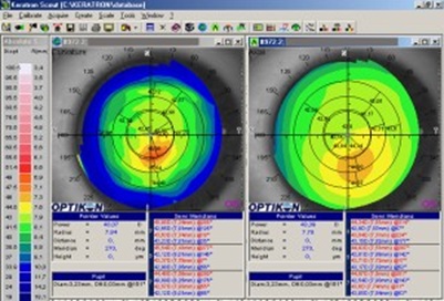
A cornea examination should include keratotopography - a study of the relief and optical power of the cornea over its entire area, optical coherent tomography, which determines the thickness, morphology and geometry of the cornea also over the entire area, the study of the back surface of the cornea using the Shimflug camera to determine the irregularities of the anterior and posterior cornea , confocal microscopy - the study of the posterior layer of the cornea (endothelium). According to the results of this survey, we can already conclude how healthy the cornea is.

This is how the examination on the Scheimflug-camera + Placido rings looks like.
Sometimes astigmatism is associated with the lens - its form, the presence of various opacities in it or a change in its position in space, such astigmatism is called lens-shaped. And it happens that the cornea is irregular and the lens is distorted, then the resultant will affect the final optics as a whole.
We can also study the anterior segment of the eye using ultrasound biomicroscopy in order to examine the parts of the eye that are inaccessible to other methods and measure them.
I described in detail how this happens in the previous post: We got to the side effects of laser vision correction - and even before the diagnosis ( fasting ) and here: “Augmentation” of the eye: what we build into it today, and what else will remain ( post ). Calculation of intraocular phakic lenses (embedded in the eye) - we continue about the eye and its biomechanics ( post ).
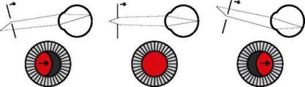
Yes, by the way, for children there is such a pediatric autorefractometer, which can measure the optics of an eye in a child, without contact, when there is no need to press anything on his forehead and everything happens at a distance of 1.5-2 m. This is important, since children in general are naughty and look badly (or refuse to look at all) wherever the ophthalmologist asks. There are not all clinics, as it is expensive. Of course, we have it, called PlusOptix. And late diagnosed astigmatism leads to serious consequences.

This is a survey with PlusOptix
And, I have not yet named the old antediluvian method of skiascopy - tracking the movement of the shadow of the reflex from the fundus with the luminescence of a reverse ophthalmoscope and moving the ruler with lenses. Our grandmothers were diagnosed this way, by the way, quite successfully, if you know how to do it.
Method skiascopy.
One of the main symptoms that appear during keratoconus is astigmatism. When we diagnose a patient with astigmatism, we must always exclude the latent (forme fruste) or the initial form of keratoconus. I wrote about this serious illness in a post:
→ Keratoectasia (keratoconus, “bulging cornea”): what it is and what to do with it
At the beginning of the disease, you may not even suspect about it at all, even astigmatism may be quite small - 0.5 - 0.75 diopters, however, changes in the cornea are already there and are available only by special methods of examination. Therefore, if you are offered to examine the cornea for its health, this is not a commercial "cheat", it is worth doing, since the treatment in the initial stages is much more effective than in developed and distant ones.
With keratoconus it is just about irregular astigmatism, which is bad in advanced stages amenable to correction.
This is the keratotopogram at the initial keratoconus.
And in the next post I will talk about how to correct astigmatism, both surgical and non-surgical, as well as what is most important in astigmatism in childhood and adulthood.
GO TO THE METHODS OF CORRECTION OF ASTIGMATISM >>>
Modern statistics depressing. More than half of the world's population suffers from visual impairment, the most common problems are myopia and hyperopia. But ophthalmologists often diagnose another disease, the name of which is unfamiliar to many. Astigmatism is a defect in the optical system of the eye when the sharpness of the image is asymmetrical in vertical and horizontal. And the parallel rays of light passing through the eye are not focused into a point, but into the “eight”. For a person, this means that the visibility of the image becomes unsharp, and this often applies to both distant and close objects. As a result, instead of a normal image, a person sees a distorted image, in which some lines are clear, others are blurred. An idea of this can be obtained if you look at your distorted reflection in an oval teaspoon. A similar distorted image is formed during astigmatism on the retina.
Ophthalmologists say that almost two thirds of the world's inhabitants face this problem. But since the degree of astigmatism may be small, many people practically do not feel any discomfort. It is difficult for doctors to identify a clear list of common symptoms indicating that a patient has astigmatism. In each case, they will vary. In the very early stages, it is often confused with tired eyes.
However, quite a lot of people need special treatment or correction of this disorder with the help of glasses, contact lenses or even surgery.
General
Astigmatism is a defect in the optical system of the eye. In this case, the refraction (or reflection) of the rays in different sections of the transmitted light beam is not the same.
As a result, the image of objects becomes blurred, each point of the object is displayed with a blurred ellipse. And the whole picture, like in a “crooked mirror”, turns into a so-called figure “Sturm's conoid”. In practice, the main complaint is blurred vision at a distance, often vague near blurring, vagueness and division of objects.
The course of the rays in the eye with astigmatism looks like.
- I was born this way or ruined my life?
This is the first question that patients ask when they hear this diagnosis for the first time.
Astigmatism may be congenital or acquired, may be stable and progress.
Quite often, astigmatism is a feature of the structure of the cornea at birth, it can even be inherited from parents. If its value affects visual acuity (as a rule, we are talking about astigmatism greater than 1 diopter), then it necessarily requires correction from an early age. Otherwise, the eye sees poorly and develops amblyopia, the “lazy” eye, which is no longer capable of high vision in adulthood. A child with astigmatism, by the way, will never complain to you about poor eyesight - he doesn’t know how to see this well. Therefore, early examinations by an oculist and wearing glasses, "as prescribed by a doctor," is so important.
It often happens that a grown-up person is surprised to find out that he has, as it turns out, astigmatism, this is in the case of those not examined in childhood.
Astigmatism may appear already in adulthood as a result of corneal diseases, such as keratoconus, inflammation, trauma and operations.
In any case, it does not appear from working at the computer.
- What is damaged in astigmatism?
First of all, the cause of astigmatism can be two optical systems of the eye - the cornea and lens. When glasses or lenses are picked, they take into account the resulting optical power of these two systems. Please note, patients usually know about frequent corneal astigmatism, but forget about the lens - it can make a difference, for example, in cataracts, when irregularities appear in the lens. "The cornea is flat, but there is astigmatism" - this is it. Often the patient and the optometrist do not bother at all with what is the cause. When selecting points, it is important to correct the rays of light onto the retina using correction. And with surgical correction - this is important.
For a simplified understanding of the essence of the problem, I often explain to patients that the optics of the eye with astigmatism is not like a sphere (a football), but an oval (a rugby ball).
- How is astigmatism different in size, shape and content?
First, the value of astigmatism matters - conditionally up to 1 diopter, astigmatism is considered physiological, weak. That is, as a rule, it does not require correction. But in case he even lowers his vision, alas, he needs to be corrected. The average degree - up to 2 diopters, high - 2-3 diopters and more than 3 diopters - this is a very high astigmatism. There are other classifications, but this one best reflects the effect on defocus.
Secondly, there are two types of corneal astigmatism: regular and irregular.
With regular astigmatism, there are two main refractive perpendicular cross-sectional planes, within which the optical power does not change - weak and strong. In such optics there is at least some regularity, even if the axes are skew or there is a big difference in their optical power.
Regular astigmatism - in this case, two parts can be distinguished, where the cornea has a different degree of refraction. This type of astigmatism can be corrected with glasses with cylindrical glasses or soft contact lenses.
Irregular astigmatism - in this case, two parts cannot be distinguished, since there are many optical axes. This type usually develops as a result of corneal damage, for example, during an accident (scar tissue forms), or due to uneven corneal protuberances (keratoconus). Irregular astigmatism is usually poorly corrected, sometimes hard contact lenses or laser effects on the cornea can be useful.
Third, the type of astigmatism depends on the location of the strong axis. If the strong axis is vertical or no more than 30 degrees deviates from the vertical - direct astigmatism. If the strong axis is horizontal ± 30 degrees - astigmatism is reversed. If in between - astigmatism with oblique axes. Direct astigmatism slightly increases the depth of focus, the reverse - more impairs visibility, but these both options are well corrected. But the correction of astigmatism with oblique axes with glasses and lenses is worse performed.
In astigmatic eyes, there are two perpendicular to the section plane with the greatest and the smallest refractive power.
Fourthly, depending on how the focus relates to the retina, astigmatism is divided into myopic (myopic), long-sighted (hypermetropic) and mixed - when one part of the image falls in front of the retina and the part behind the retina. For example, minus two and minus four - then this is complex myopic astigmatism, plus two and plus four - complex long-sighted astigmatism, plus two and minus four - mixed.
Nearsighted astigmatism is indicated by the sign “-”, long-sighted - by the sign “+”, when mixed in the eye there are “+” and “-”.
- How can I suspect astigmatism?
Symptoms of astigmatism are a decrease in vision, sometimes the vision of objects is bent, their split, rapid eyestrain at work, headache. Often you may not even suspect that there is such a problem, complaints are so typical - poor vision for distance or near, or at all distances.
- What kind of diagnosis is needed for astigmatism.
Since astigmatism is a defect in optics, then, firstly, the optics of the eye are carefully examined. And the correct conclusion will give only an ophthalmologist. To do this, we have a series of tests: a study on an auto-refractkeratometer, an aberrometer, a phoropterer with the selection of test lenses to determine visual acuity with maximum correction. A very interesting radiant figure gives an idea of how much astigmatism is present - if you look at the center of the figure from below, some of the lines will be clearer, and some will "blur" with those who have astigmatism.
In the phoropter tests, an ophthalmologist in the arsenal also has a number of techniques to clarify the presence of astigmatism, its axis and size. And this process is always quite time consuming - this can be confirmed by those patients who have done with us, for example, SMILE. Verification of tests with "fogging, cross-cylinders with power and axial samples" for a time period of 20-30 minutes.
So, when it is determined that there is still astigmatism, it is necessary to determine "who is to blame" - the cornea or the lens.
A cornea examination should include keratotopography - a study of the relief and optical power of the cornea over its entire area, optical coherent tomography, which determines the thickness, morphology and geometry of the cornea also over the entire area, the study of the back surface of the cornea using the Shimflug camera to determine the irregularities of the anterior and posterior cornea , confocal microscopy - the study of the posterior layer of the cornea (endothelium). According to the results of this survey, we can already conclude how healthy the cornea is.
This is how the examination on the Scheimflug-camera + Placido rings looks like.
Sometimes astigmatism is associated with the lens - its form, the presence of various opacities in it or a change in its position in space, such astigmatism is called lens-shaped. And it happens that the cornea is irregular and the lens is distorted, then the resultant will affect the final optics as a whole.
We can also study the anterior segment of the eye using ultrasound biomicroscopy in order to examine the parts of the eye that are inaccessible to other methods and measure them.
I described in detail how this happens in the previous post: We got to the side effects of laser vision correction - and even before the diagnosis ( fasting ) and here: “Augmentation” of the eye: what we build into it today, and what else will remain ( post ). Calculation of intraocular phakic lenses (embedded in the eye) - we continue about the eye and its biomechanics ( post ).
Yes, by the way, for children there is such a pediatric autorefractometer, which can measure the optics of an eye in a child, without contact, when there is no need to press anything on his forehead and everything happens at a distance of 1.5-2 m. This is important, since children in general are naughty and look badly (or refuse to look at all) wherever the ophthalmologist asks. There are not all clinics, as it is expensive. Of course, we have it, called PlusOptix. And late diagnosed astigmatism leads to serious consequences.
This is a survey with PlusOptix
And, I have not yet named the old antediluvian method of skiascopy - tracking the movement of the shadow of the reflex from the fundus with the luminescence of a reverse ophthalmoscope and moving the ruler with lenses. Our grandmothers were diagnosed this way, by the way, quite successfully, if you know how to do it.
Method skiascopy.
- How is astigmatism associated with keratoconus
One of the main symptoms that appear during keratoconus is astigmatism. When we diagnose a patient with astigmatism, we must always exclude the latent (forme fruste) or the initial form of keratoconus. I wrote about this serious illness in a post:
→ Keratoectasia (keratoconus, “bulging cornea”): what it is and what to do with it
At the beginning of the disease, you may not even suspect about it at all, even astigmatism may be quite small - 0.5 - 0.75 diopters, however, changes in the cornea are already there and are available only by special methods of examination. Therefore, if you are offered to examine the cornea for its health, this is not a commercial "cheat", it is worth doing, since the treatment in the initial stages is much more effective than in developed and distant ones.
With keratoconus it is just about irregular astigmatism, which is bad in advanced stages amenable to correction.
This is the keratotopogram at the initial keratoconus.
And in the next post I will talk about how to correct astigmatism, both surgical and non-surgical, as well as what is most important in astigmatism in childhood and adulthood.
GO TO THE METHODS OF CORRECTION OF ASTIGMATISM >>>
All Articles